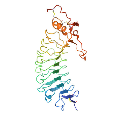
1GWB

STRUCTURE OF GLYCOPROTEIN 1B


Experimental Data Snapshot

  • Method: X-RAY DIFFRACTION
  • Resolution: 2.80 Å
  • R-Value Free: 0.274 
  • R-Value Work: 0.245 
  • R-Value Observed: 0.245 

wwPDB Validation   3D Report Full Report


Ligand Structure Quality Assessment 


This is version 1.5 of the entry. See complete history


Literature

Crystal Structure of the Platelet Glycoprotein Ib-Alpha N-Terminal Domain Reveals an Unmasking Mechanism of Receptor Activation

Uff, S.Clemetson, J.M.Harrison, T.Clemetson, K.J.Emsley, J.

(2002) J Biol Chem 277: 35657

  • DOI: https://doi.org/10.1074/jbc.M205271200
  • Primary Citation of Related Structures:  
    1GWB

  • PubMed Abstract: 

    Glycoprotein Ib (GPIb) is a platelet receptor with a critical role in mediating the arrest of platelets at sites of vascular damage. GPIb binds to the A1 domain of von Willebrand factor (vWF-A1) at high blood shear, initiating platelet adhesion and contributing to the formation of a thrombus. To investigate the molecular basis of GPIb regulation and ligand binding, we have determined the structure of the N-terminal domain of the GPIb(alpha) chain (residues 1-279). This structure is the first determined from the cell adhesion/signaling class of leucine-rich repeat (LRR) proteins and reveals the topology of the characteristic disulfide-bonded flanking regions. The fold consists of an N-terminal beta-hairpin, eight leucine-rich repeats, a disulfide-bonded loop, and a C-terminal anionic region. The structure also demonstrates a novel LRR motif in the form of an M-shaped arrangement of three tandem beta-turns. Negatively charged binding surfaces on the LRR concave face and anionic region indicate two-step binding kinetics to vWF-A1, which can be regulated by an unmasking mechanism involving conformational change of a key loop. Using molecular docking of the GPIb and vWF-A1 crystal structures, we were also able to model the GPIb.vWF-A1 complex.


  • Organizational Affiliation

    Department of Biochemistry, University of Leicester, Leicester LE1 7RH, United Kingdom.


Macromolecules
Find similar proteins by:  (by identity cutoff)  |  3D Structure
Entity ID: 1
MoleculeChains Sequence LengthOrganismDetailsImage
PLATELET GLYCOPROTEIN IB ALPHA CHAIN
A, B
281Homo sapiensMutation(s): 0 
UniProt & NIH Common Fund Data Resources
Find proteins for P07359 (Homo sapiens)
Explore P07359 
Go to UniProtKB:  P07359
PHAROS:  P07359
GTEx:  ENSG00000185245 
Entity Groups  
Sequence Clusters30% Identity50% Identity70% Identity90% Identity95% Identity100% Identity
UniProt GroupP07359
Glycosylation
Glycosylation Sites: 1Go to GlyGen: P07359-1
Sequence Annotations
Expand
  • Reference Sequence
Small Molecules
Ligands 4 Unique
IDChains Name / Formula / InChI Key2D Diagram3D Interactions
NAG
Query on NAG

Download Ideal Coordinates CCD File 
F [auth A],
G [auth A],
J [auth B]
2-acetamido-2-deoxy-beta-D-glucopyranose
C8 H15 N O6
OVRNDRQMDRJTHS-FMDGEEDCSA-N
PT
Query on PT

Download Ideal Coordinates CCD File 
C [auth A],
D [auth A],
H [auth B],
I [auth B]
PLATINUM (II) ION
Pt
HRGDZIGMBDGFTC-UHFFFAOYSA-N
SO4
Query on SO4

Download Ideal Coordinates CCD File 
K [auth B]SULFATE ION
O4 S
QAOWNCQODCNURD-UHFFFAOYSA-L
ACY
Query on ACY

Download Ideal Coordinates CCD File 
E [auth A]ACETIC ACID
C2 H4 O2
QTBSBXVTEAMEQO-UHFFFAOYSA-N
Modified Residues  1 Unique
IDChains TypeFormula2D DiagramParent
TYS
Query on TYS
A, B
L-PEPTIDE LINKINGC9 H11 N O6 STYR
Experimental Data & Validation

Experimental Data

  • Method: X-RAY DIFFRACTION
  • Resolution: 2.80 Å
  • R-Value Free: 0.274 
  • R-Value Work: 0.245 
  • R-Value Observed: 0.245 
  • Space Group: P 64 2 2
Unit Cell:
Length ( Å )Angle ( ˚ )
a = 202α = 90
b = 202β = 90
c = 128γ = 120
Software Package:
Software NamePurpose
CNSrefinement

Structure Validation

View Full Validation Report


Ligand Structure Quality Assessment 


Entry History 

Deposition Data

Revision History  (Full details and data files)

  • Version 1.0: 2003-02-06
    Type: Initial release
  • Version 1.1: 2011-10-19
    Changes: Advisory, Atomic model, Derived calculations, Non-polymer description, Other, Refinement description, Source and taxonomy, Structure summary, Version format compliance
  • Version 1.2: 2012-01-18
    Changes: Other
  • Version 1.3: 2017-07-12
    Changes: Advisory
  • Version 1.4: 2020-07-29
    Type: Remediation
    Reason: Carbohydrate remediation
    Changes: Data collection, Derived calculations, Other, Structure summary
  • Version 1.5: 2024-11-13
    Changes: Data collection, Database references, Structure summary