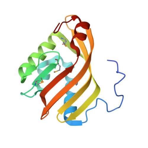
1NU3

Limonene-1,2-epoxide hydrolase in complex with valpromide


Experimental Data Snapshot

  • Method: X-RAY DIFFRACTION
  • Resolution: 1.75 Å
  • R-Value Free: 0.195 
  • R-Value Work: 0.158 
  • R-Value Observed: 0.159 

wwPDB Validation   3D Report Full Report


This is version 1.5 of the entry. See complete history


Literature

Structure of Rhodococcus erythropolis limonene-1,2-epoxide hydrolase reveals a novel active site

Arand, M.Hallberg, B.M.Zou, J.Bergfors, T.Oesch, F.van der Werf, M.J.de Bont, J.A.M.Jones, T.A.Mowbray, S.L.

(2003) EMBO J 22: 2583-2592

  • DOI: https://doi.org/10.1093/emboj/cdg275
  • Primary Citation of Related Structures:  
    1NU3, 1NWW

  • PubMed Abstract: 

    Epoxide hydrolases are essential for the processing of epoxide-containing compounds in detoxification or metabolism. The classic epoxide hydrolases have an alpha/beta hydrolase fold and act via a two-step reaction mechanism including an enzyme-substrate intermediate. We report here the structure of the limonene-1,2-epoxide hydrolase from Rhodococcus erythropolis, solved using single-wavelength anomalous dispersion from a selenomethionine-substituted protein and refined at 1.2 A resolution. This enzyme represents a completely different structure and a novel one-step mechanism. The fold features a highly curved six-stranded mixed beta-sheet, with four alpha-helices packed onto it to create a deep pocket. Although most residues lining this pocket are hydrophobic, a cluster of polar groups, including an Asp-Arg-Asp triad, interact at its deepest point. Site-directed mutagenesis supports the conclusion that this is the active site. Further, a 1.7 A resolution structure shows the inhibitor valpromide bound at this position, with its polar atoms interacting directly with the residues of the triad. We suggest that several bacterial proteins of currently unknown function will share this structure and, in some cases, catalytic properties.


  • Organizational Affiliation

    Department of Toxicology, University of Würzburg, Versbacher Strasse 9, D-97078 Würzburg, Germany.


Macromolecules
Find similar proteins by:  (by identity cutoff)  |  3D Structure
Entity ID: 1
MoleculeChains Sequence LengthOrganismDetailsImage
limonene-1,2-epoxide hydrolase
A, B
149Rhodococcus erythropolisMutation(s): 5 
Gene Names: limA
EC: 3.3.2.8
UniProt
Find proteins for Q9ZAG3 (Rhodococcus erythropolis)
Explore Q9ZAG3 
Go to UniProtKB:  Q9ZAG3
Entity Groups  
Sequence Clusters30% Identity50% Identity70% Identity90% Identity95% Identity100% Identity
UniProt GroupQ9ZAG3
Sequence Annotations
Expand
  • Reference Sequence
Small Molecules
Modified Residues  1 Unique
IDChains TypeFormula2D DiagramParent
MSE
Query on MSE
A, B
L-PEPTIDE LINKINGC5 H11 N O2 SeMET
Binding Affinity Annotations 
IDSourceBinding Affinity
VPR PDBBind:  1NU3 Ki: 1.00e+5 (nM) from 1 assay(s)
Experimental Data & Validation

Experimental Data

  • Method: X-RAY DIFFRACTION
  • Resolution: 1.75 Å
  • R-Value Free: 0.195 
  • R-Value Work: 0.158 
  • R-Value Observed: 0.159 
  • Space Group: P 21 21 21
Unit Cell:
Length ( Å )Angle ( ˚ )
a = 45.596α = 90
b = 47.656β = 90
c = 129.244γ = 90
Software Package:
Software NamePurpose
REFMACrefinement
MOSFLMdata reduction
CCP4data scaling
FFTphasing

Structure Validation

View Full Validation Report


Entry History 

Deposition Data

Revision History  (Full details and data files)

  • Version 1.0: 2003-06-10
    Type: Initial release
  • Version 1.1: 2008-04-29
    Changes: Version format compliance
  • Version 1.2: 2011-07-13
    Changes: Version format compliance
  • Version 1.3: 2018-05-23
    Changes: Data collection
  • Version 1.4: 2021-11-10
    Changes: Database references, Derived calculations
  • Version 1.5: 2024-11-20
    Changes: Data collection, Structure summary