2G83

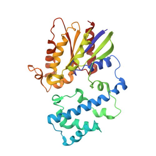
Structure of activated G-alpha-i1 bound to a nucleotide-state-selective peptide: Minimal determinants for recognizing the active form of a G protein alpha subunit


Experimental Data Snapshot

  • Method: X-RAY DIFFRACTION
  • Resolution: 2.80 Å
  • R-Value Free: 0.300 
  • R-Value Work: 0.269 
  • R-Value Observed: 0.301 

Starting Model: experimental
View more details

wwPDB Validation   3D Report Full Report


Ligand Structure Quality Assessment 


This is version 1.4 of the entry. See complete history


Literature

Minimal Determinants for Binding Activated Galpha from the Structure of a Galpha(i1)-Peptide Dimer.

Johnston, C.A.Lobanova, E.S.Shavkunov, A.S.Low, J.Ramer, J.K.Blaesius, R.Fredericks, Z.Willard, F.S.Kuhlman, B.Arshavsky, V.Y.Siderovski, D.P.

(2006) Biochemistry 45: 11390-11400

  • DOI: https://doi.org/10.1021/bi0613832
  • Primary Citation of Related Structures:  
    2G83

  • PubMed Abstract: 

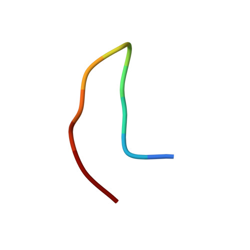
    G-proteins cycle between an inactive GDP-bound state and an active GTP-bound state, serving as molecular switches that coordinate cellular signaling. We recently used phage display to identify a series of peptides that bind G alpha subunits in a nucleotide-dependent manner [Johnston, C. A., Willard, F. S., Jezyk, M. R., Fredericks, Z., Bodor, E. T., Jones, M. B., Blaesius, R., Watts, V. J., Harden, T. K., Sondek, J., Ramer, J. K., and Siderovski, D. P. (2005) Structure 13, 1069-1080]. Here we describe the structural features and functions of KB-1753, a peptide that binds selectively to GDP x AlF4(-)- and GTPgammaS-bound states of G alpha(i) subunits. KB-1753 blocks interaction of G alpha(transducin) with its effector, cGMP phosphodiesterase, and inhibits transducin-mediated activation of cGMP degradation. Additionally, KB-1753 interferes with RGS protein binding and resultant GAP activity. A fluorescent KB-1753 variant was found to act as a sensor for activated G alpha in vitro. The crystal structure of KB-1753 bound to G alpha(i1) x GDP x AlF4(-) reveals binding to a conserved hydrophobic groove between switch II and alpha3 helices and, along with supporting biochemical data and previous structural analyses, supports the notion that this is the site of effector interactions for G alpha(i) subunits.


  • Organizational Affiliation

    Department of Pharmacology, University of North Carolina School of Medicine, Chapel Hill, North Carolina 27599-7365, USA.


Macromolecules
Find similar proteins by:  (by identity cutoff)  |  3D Structure
Entity ID: 1
MoleculeChains Sequence LengthOrganismDetailsImage
Guanine nucleotide-binding protein G(i), alpha-1 subunit
A, B
313Homo sapiensMutation(s): 0 
Gene Names: GNAI1
EC: 3.6.5
UniProt & NIH Common Fund Data Resources
Find proteins for P63096 (Homo sapiens)
Explore P63096 
Go to UniProtKB:  P63096
PHAROS:  P63096
GTEx:  ENSG00000127955 
Entity Groups  
Sequence Clusters30% Identity50% Identity70% Identity90% Identity95% Identity100% Identity
UniProt GroupP63096
Sequence Annotations
Expand
  • Reference Sequence

Find similar proteins by:  Sequence   |   3D Structure  

Entity ID: 2
MoleculeChains Sequence LengthOrganismDetailsImage
KB-1753 phage display peptide
C, D
11N/AMutation(s): 0 
Entity Groups  
Sequence Clusters30% Identity50% Identity70% Identity90% Identity95% Identity100% Identity
Sequence Annotations
Expand
  • Reference Sequence
Small Molecules
Binding Affinity Annotations 
IDSourceBinding Affinity
GDP PDBBind:  2G83 Kd: 1200 (nM) from 1 assay(s)
Experimental Data & Validation

Experimental Data

  • Method: X-RAY DIFFRACTION
  • Resolution: 2.80 Å
  • R-Value Free: 0.300 
  • R-Value Work: 0.269 
  • R-Value Observed: 0.301 
  • Space Group: P 32 2 1
Unit Cell:
Length ( Å )Angle ( ˚ )
a = 103.13α = 90
b = 103.13β = 90
c = 206.99γ = 120
Software Package:
Software NamePurpose
CNSrefinement
PDB_EXTRACTdata extraction
HKL-2000data reduction
HKL-2000data scaling
AMoREphasing

Structure Validation

View Full Validation Report


Ligand Structure Quality Assessment 


Entry History 

Deposition Data

Revision History  (Full details and data files)

  • Version 1.0: 2006-10-10
    Type: Initial release
  • Version 1.1: 2008-05-01
    Changes: Version format compliance
  • Version 1.2: 2011-07-13
    Changes: Version format compliance
  • Version 1.3: 2023-08-30
    Changes: Data collection, Database references, Derived calculations, Refinement description
  • Version 1.4: 2024-12-25
    Changes: Advisory, Derived calculations, Structure summary