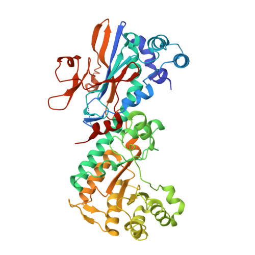
2G97

Crystal Structure of Visfatin/Pre-B Cell Colony Enhancing Factor 1/Nicotinamide Phosphoribosyltransferase In Complex with the Specific Inhibitor FK-866


Experimental Data Snapshot

  • Method: X-RAY DIFFRACTION
  • Resolution: 2.90 Å
  • R-Value Free: 0.268 
  • R-Value Work: 0.240 
  • R-Value Observed: 0.243 

Starting Model: experimental
View more details

wwPDB Validation   3D Report Full Report


Ligand Structure Quality Assessment 


This is version 1.4 of the entry. See complete history


Literature

Crystal Structure of Visfatin/Pre-B Cell Colony-enhancing Factor 1/Nicotinamide Phosphoribosyltransferase, Free and in Complex with the Anti-cancer Agent FK-866

Kim, M.-K.Lee, J.H.Kim, H.Park, S.J.Kim, S.H.Kang, G.B.Lee, Y.S.Kim, J.B.Kim, K.K.Suh, S.W.Eom, S.H.

(2006) J Mol Biol 362: 66-77

  • DOI: https://doi.org/10.1016/j.jmb.2006.06.082
  • Primary Citation of Related Structures:  
    2G95, 2G96, 2G97

  • PubMed Abstract: 

    Visfatin/pre-B cell colony-enhancing factor 1 (PBEF)/nicotinamide phosphoribosyltransferase (NAmPRTase) is a multifunctional protein having phosphoribosyltransferase, cytokine and adipokine activities. Originally isolated as a cytokine promoting the differentiation of B cell precursors, it was recently suggested to act as an insulin analog via the insulin receptor. Here, we describe the first crystal structure of visfatin in three different forms: apo and in complex with either nicotinamide mononucleotide (NMN) or the NAmPRTase inhibitor FK-866 which was developed as an anti-cancer agent, interferes with NAD biosynthesis, showing a particularly high specificity for NAmPRTase. The crystal structures of the complexes with either NMN or FK-866 show that the enzymatic active site of visfatin is optimized for nicotinamide binding and that the nicotinamide-binding site is important for inhibition by FK-866. Interestingly, visfatin mimics insulin signaling by binding to the insulin receptor with an affinity similar to that of insulin and does not share the binding site with insulin on the insulin receptor. To predict binding sites, the potential interaction patches of visfatin and the L1-CR-L2 domain of insulin receptor were generated and analyzed. Although the relationship between the insulin-mimetic property and the enzymatic function of visfatin has not been clearly established, our structures raise the intriguing possibility that the glucose metabolism and the NAD biosynthesis are linked by visfatin.


  • Organizational Affiliation

    Department of Life Science, Gwangju Institute of Science & Technology, Gwangju 500-712, Korea.


Macromolecules
Find similar proteins by:  (by identity cutoff)  |  3D Structure
Entity ID: 1
MoleculeChains Sequence LengthOrganismDetailsImage
Nicotinamide phosphoribosyltransferase
A, B
491Rattus norvegicusMutation(s): 0 
EC: 2.4.2.12
UniProt
Find proteins for Q80Z29 (Rattus norvegicus)
Explore Q80Z29 
Go to UniProtKB:  Q80Z29
Entity Groups  
Sequence Clusters30% Identity50% Identity70% Identity90% Identity95% Identity100% Identity
UniProt GroupQ80Z29
Sequence Annotations
Expand
  • Reference Sequence
Small Molecules
Ligands 1 Unique
IDChains Name / Formula / InChI Key2D Diagram3D Interactions
DGB
Query on DGB

Download Ideal Coordinates CCD File 
C [auth A],
D [auth B]
(2E)-N-{4-[1-(benzenecarbonyl)piperidin-4-yl]butyl}-3-(pyridin-3-yl)prop-2-enamide
C24 H29 N3 O2
KPBNHDGDUADAGP-VAWYXSNFSA-N
Binding Affinity Annotations 
IDSourceBinding Affinity
DGB BindingDB:  2G97 Ki: min: 0.3, max: 3 (nM) from 2 assay(s)
Kd: 10 (nM) from 1 assay(s)
IC50: min: 0.14, max: 35 (nM) from 17 assay(s)
PDBBind:  2G97 Kd: 152 (nM) from 1 assay(s)
Experimental Data & Validation

Experimental Data

  • Method: X-RAY DIFFRACTION
  • Resolution: 2.90 Å
  • R-Value Free: 0.268 
  • R-Value Work: 0.240 
  • R-Value Observed: 0.243 
  • Space Group: P 21 21 21
Unit Cell:
Length ( Å )Angle ( ˚ )
a = 82.021α = 90
b = 106.482β = 90
c = 118.061γ = 90
Software Package:
Software NamePurpose
HKL-2000data collection
SCALEPACKdata scaling
CNSrefinement
HKL-2000data reduction
CNSphasing

Structure Validation

View Full Validation Report


Ligand Structure Quality Assessment 


Entry History 

Deposition Data

Revision History  (Full details and data files)

  • Version 1.0: 2006-08-01
    Type: Initial release
  • Version 1.1: 2008-05-01
    Changes: Version format compliance
  • Version 1.2: 2011-07-13
    Changes: Version format compliance
  • Version 1.3: 2016-11-09
    Changes: Non-polymer description
  • Version 1.4: 2023-10-25
    Changes: Data collection, Database references, Derived calculations, Refinement description