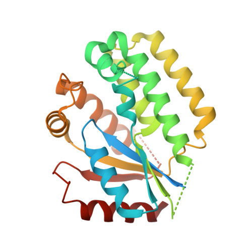
2ZI7

C4S dCK variant of dCK in complex with D-dG+UDP


Experimental Data Snapshot

  • Method: X-RAY DIFFRACTION
  • Resolution: 1.97 Å
  • R-Value Free: 0.259 
  • R-Value Work: 0.206 
  • R-Value Observed: 0.211 

Starting Model: experimental
View more details

wwPDB Validation   3D Report Full Report


Ligand Structure Quality Assessment 


This is version 1.4 of the entry. See complete history


Literature

Elucidation of different binding modes of purine nucleosides to human deoxycytidine kinase

Sabini, E.Hazra, S.Konrad, M.Lavie, A.

(2008) J Med Chem 51: 4219-4225

  • DOI: https://doi.org/10.1021/jm800134t
  • Primary Citation of Related Structures:  
    2ZI7, 2ZI9, 2ZIA

  • PubMed Abstract: 

    Purine nucleoside analogues of medicinal importance, such as cladribine, require phosphorylation by deoxycytidine kinase (dCK) for pharmacological activity. Structural studies of ternary complexes of human dCK show that the enzyme conformation adjusts to the different hydrogen-bonding properties between dA and dG and to the presence of substituent at the 2-position present in dG and cladribine. Specifically, the carbonyl group in dG elicits a previously unseen conformational adjustment of the active site residues Arg104 and Asp133. In addition, dG and cladribine adopt the anti conformation, in contrast to the syn conformation observed with dA. Kinetic analysis reveals that cladribine is phosphorylated at the highest efficiency with UTP as donor. We attribute this to the ability of cladribine to combine advantageous properties from dA (favorable hydrogen-bonding pattern) and dG (propensity to bind to the enzyme in its anti conformation), suggesting that dA analogues with a substituent at the 2-position are likely to be better activated by human dCK.


  • Organizational Affiliation

    Department of Biochemistry and Molecular Genetics, University of Illinois at Chicago, 900 S. Ashland (M/C 669), Chicago, Illinois 60607, USA.


Macromolecules
Find similar proteins by:  (by identity cutoff)  |  3D Structure
Entity ID: 1
MoleculeChains Sequence LengthOrganismDetailsImage
Deoxycytidine kinaseA [auth B],
B [auth A]
279Homo sapiensMutation(s): 4 
Gene Names: DCK
EC: 2.7.1.74 (PDB Primary Data), 2.7.1.113 (UniProt), 2.7.1.76 (UniProt)
UniProt & NIH Common Fund Data Resources
Find proteins for P27707 (Homo sapiens)
Explore P27707 
Go to UniProtKB:  P27707
PHAROS:  P27707
GTEx:  ENSG00000156136 
Entity Groups  
Sequence Clusters30% Identity50% Identity70% Identity90% Identity95% Identity100% Identity
UniProt GroupP27707
Sequence Annotations
Expand
  • Reference Sequence
Experimental Data & Validation

Experimental Data

  • Method: X-RAY DIFFRACTION
  • Resolution: 1.97 Å
  • R-Value Free: 0.259 
  • R-Value Work: 0.206 
  • R-Value Observed: 0.211 
  • Space Group: P 21 21 21
Unit Cell:
Length ( Å )Angle ( ˚ )
a = 43.8α = 90
b = 107.5β = 90
c = 110.4γ = 90
Software Package:
Software NamePurpose
REFMACrefinement
SERGUIdata collection
XDSdata reduction
XDSdata scaling
MOLREPphasing

Structure Validation

View Full Validation Report


Ligand Structure Quality Assessment 


Entry History 

Deposition Data

Revision History  (Full details and data files)

  • Version 1.0: 2008-07-08
    Type: Initial release
  • Version 1.1: 2011-07-13
    Changes: Version format compliance
  • Version 1.2: 2021-11-10
    Changes: Database references, Derived calculations
  • Version 1.3: 2023-11-01
    Changes: Data collection, Refinement description
  • Version 1.4: 2024-12-25
    Changes: Advisory, Derived calculations, Structure summary