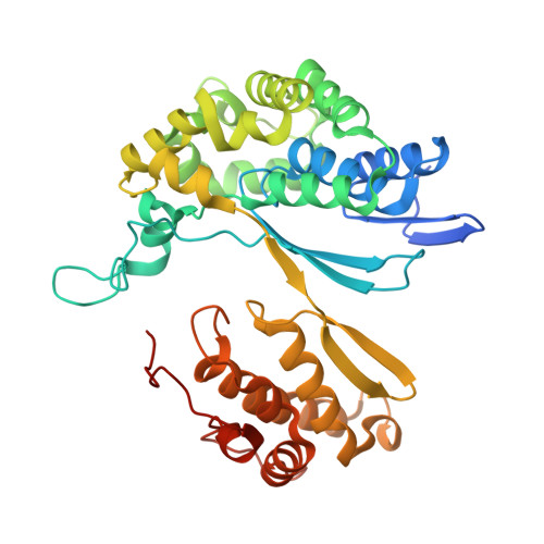
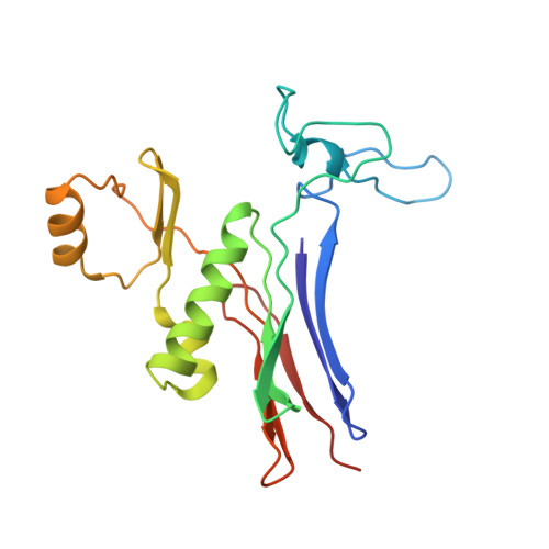
3A75

Crystal structure of glutamate complex of halotolerant γ-glutamyltranspeptidase from Bacillus subtilis


Experimental Data Snapshot

  • Method: X-RAY DIFFRACTION
  • Resolution: 1.95 Å
  • R-Value Free: 0.262 
  • R-Value Work: 0.208 
  • R-Value Observed: 0.208 

Starting Model: experimental
View more details

wwPDB Validation   3D Report Full Report


This is version 1.2 of the entry. See complete history


Literature

Crystal structure of the halotolerant gamma-glutamyltranspeptidase from Bacillus subtilis in complex with glutamate reveals a unique architecture of the solvent-exposed catalytic pocket

Wada, K.Irie, M.Suzuki, H.Fukuyama, K.

(2010) FEBS J 277: 1000-1009

  • DOI: https://doi.org/10.1111/j.1742-4658.2009.07543.x
  • Primary Citation of Related Structures:  
    3A75

  • PubMed Abstract: 

    gamma-Glutamyltranspeptidase (GGT; EC 2.3.2.2), an enzyme found in organisms from bacteria to mammals and plants, plays a central role in glutathione metabolism. Structural studies of GGTs from Escherichia coli and Helicobacter pylori have revealed detailed molecular mechanisms of catalysis and maturation. In these two GGTs, highly conserved residues form the catalytic pockets, conferring the ability of the loop segment to shield the bound gamma-glutamyl moiety from the solvent. Here, we have examined the Bacillus subtilis GGT, which apparently lacks the amino acids corresponding to the lid-loop that are present in mammalian and plant GGTs as well as in most bacterial GGTs. Another remarkable feature of B. subtilis GGT is its salt tolerance; it retains 86% of its activity even in 3 m NaCl. To better understand these characteristics of B. subtilis GGT, we determined its crystal structure in complex with glutamate, a product of the enzymatic reaction, at 1.95 A resolution. This structure revealed that, unlike the E. coli and H. pylori GGTs, the catalytic pocket of B. subtilis GGT has no segment that covers the bound glutamate; consequently, the glutamate is exposed to solvent. Furthermore, calculation of the electrostatic potential showed that strong acidic patches were distributed on the surface of the B. subtilis GGT, even under high-salt conditions, and this may allow the protein to remain in the hydrated state and avoid self-aggregation. The structural findings presented here have implications for the molecular mechanism of GGT.


  • Organizational Affiliation

    Department of Biological Sciences, Graduate School of Science, Osaka University, Japan.


Macromolecules
Find similar proteins by:  (by identity cutoff)  |  3D Structure
Entity ID: 1
MoleculeChains Sequence LengthOrganismDetailsImage
Gamma-glutamyltranspeptidase large chain
A, C
384Bacillus subtilisMutation(s): 0 
EC: 2.3.2.2 (PDB Primary Data), 3.4.19.13 (UniProt)
UniProt
Find proteins for P54422 (Bacillus subtilis (strain 168))
Explore P54422 
Go to UniProtKB:  P54422
Entity Groups  
Sequence Clusters30% Identity50% Identity70% Identity90% Identity95% Identity100% Identity
UniProt GroupP54422
Sequence Annotations
Expand
  • Reference Sequence
Find similar proteins by:  (by identity cutoff)  |  3D Structure
Entity ID: 2
MoleculeChains Sequence LengthOrganismDetailsImage
Gamma-glutamyltranspeptidase small chain
B, D
185Bacillus subtilisMutation(s): 0 
EC: 2.3.2.2 (PDB Primary Data), 3.4.19.13 (UniProt)
UniProt
Find proteins for P54422 (Bacillus subtilis (strain 168))
Explore P54422 
Go to UniProtKB:  P54422
Entity Groups  
Sequence Clusters30% Identity50% Identity70% Identity90% Identity95% Identity100% Identity
UniProt GroupP54422
Sequence Annotations
Expand
  • Reference Sequence
Experimental Data & Validation

Experimental Data

  • Method: X-RAY DIFFRACTION
  • Resolution: 1.95 Å
  • R-Value Free: 0.262 
  • R-Value Work: 0.208 
  • R-Value Observed: 0.208 
  • Space Group: P 21 21 21
Unit Cell:
Length ( Å )Angle ( ˚ )
a = 49.4α = 90
b = 98.89β = 90
c = 227.2γ = 90
Software Package:
Software NamePurpose
BSSdata collection
MOLREPphasing
CNSrefinement
XDSdata reduction
XDSdata scaling

Structure Validation

View Full Validation Report


Entry History 

Deposition Data

Revision History  (Full details and data files)

  • Version 1.0: 2010-03-09
    Type: Initial release
  • Version 1.1: 2011-07-13
    Changes: Version format compliance
  • Version 1.2: 2023-11-01
    Changes: Data collection, Database references, Derived calculations, Refinement description