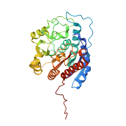
3KV2 | pdb_00003kv2

HIGH RESOLUTION STRUCTURE OF HUMAN ARGINASE I IN COMPLEX WITH THE STRONG INHIBITOR N(omega)-hydroxy-nor-L-arginine (nor-NOHA)


Experimental Data Snapshot

  • Method: X-RAY DIFFRACTION
  • Resolution: 1.55 Å
  • R-Value Free: 
    0.178 (Depositor), 0.150 (DCC) 
  • R-Value Work: 
    0.144 (Depositor), 0.150 (DCC) 
  • R-Value Observed: 
    0.144 (Depositor) 

Starting Model: experimental
View more details

wwPDB Validation   3D Report Full Report


Ligand Structure Quality Assessment 

Created with Raphaël 2.3.0Worse 01 BetterLigand structure goodness of fit to experimental dataBest fitted NNHClick on this verticalbar to view details

This is version 1.2 of the entry. See complete history


Literature

Inhibition of human arginase I by substrate and product analogues.

Di Costanzo, L.Ilies, M.Thorn, K.J.Christianson, D.W.

(2010) Arch Biochem Biophys 496: 101-108

  • DOI: https://doi.org/10.1016/j.abb.2010.02.004
  • Primary Citation of Related Structures:  
    3KV2, 3LP4, 3LP7

  • PubMed Abstract: 

    Human arginase I is a binuclear manganese metalloenzyme that catalyzes the hydrolysis of L-arginine to generate L-ornithine and urea. We demonstrate that N-hydroxy-L-arginine (NOHA) binds to this enzyme with K(d)=3.6 microM, and nor-N-hydroxy-L-arginine (nor-NOHA) binds with K(d)=517 nM (surface plasmon resonance) or K(d) approximately 50 nM (isothermal titration calorimetry). Crystals of human arginase I complexed with NOHA and nor-NOHA afford 2.04 and 1.55 A resolution structures, respectively, which are significantly improved in comparison with previously-determined structures of the corresponding complexes with rat arginase I. Higher resolution structures clarify the binding interactions of the inhibitors. Finally, the crystal structure of the complex with L-lysine (K(d)=13 microM) is reported at 1.90 A resolution. This structure confirms the importance of hydrogen bond interactions with inhibitor alpha-carboxylate and alpha-amino groups as key specificity determinants of amino acid recognition in the arginase active site.


  • Organizational Affiliation

    Roy and Diana Vagelos Laboratories, Department of Chemistry, University of Pennsylvania, Philadelphia, PA 19104-6323, USA.


Macromolecules
Find similar proteins by:  (by identity cutoff)  |  3D Structure
Entity ID: 1
MoleculeChains Sequence LengthOrganismDetailsImage
Arginase-1
A, B
322Homo sapiensMutation(s): 0 
Gene Names: ARG1
EC: 3.5.3.1
UniProt & NIH Common Fund Data Resources
Find proteins for P05089 (Homo sapiens)
Explore P05089 
Go to UniProtKB:  P05089
PHAROS:  P05089
GTEx:  ENSG00000118520 
Entity Groups  
Sequence Clusters30% Identity50% Identity70% Identity90% Identity95% Identity100% Identity
UniProt GroupP05089
Sequence Annotations
Expand
  • Reference Sequence
Small Molecules
Binding Affinity Annotations 
IDSourceBinding Affinity
NNH BindingDB:  3KV2 Ki: 500 (nM) from 1 assay(s)
Kd: min: 47, max: 517 (nM) from 3 assay(s)
IC50: min: 450, max: 5.00e+4 (nM) from 10 assay(s)
Experimental Data & Validation

Experimental Data

  • Method: X-RAY DIFFRACTION
  • Resolution: 1.55 Å
  • R-Value Free:  0.178 (Depositor), 0.150 (DCC) 
  • R-Value Work:  0.144 (Depositor), 0.150 (DCC) 
  • R-Value Observed: 0.144 (Depositor) 
Space Group: P 3
Unit Cell:
Length ( Å )Angle ( ˚ )
a = 90.497α = 90
b = 90.497β = 90
c = 69.604γ = 120
Software Package:
Software NamePurpose
CNSrefinement
HKL-2000data reduction
SCALEPACKdata scaling

Structure Validation

View Full Validation Report


Ligand Structure Quality Assessment 

Created with Raphaël 2.3.0Worse 01 BetterLigand structure goodness of fit to experimental dataBest fitted NNHClick on this verticalbar to view details

Entry History 

Deposition Data

Revision History  (Full details and data files)

  • Version 1.0: 2009-12-22
    Type: Initial release
  • Version 1.1: 2011-07-13
    Changes: Version format compliance
  • Version 1.2: 2023-09-06
    Changes: Data collection, Database references, Derived calculations, Refinement description