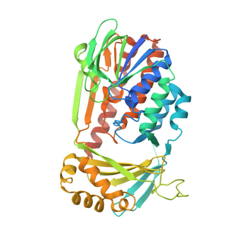
Structural basis of kynurenine 3-monooxygenase inhibition.
Amaral, M., Levy, C., Heyes, D.J., Lafite, P., Outeiro, T.F., Giorgini, F., Leys, D., Scrutton, N.S.(2013) Nature 496: 382-385
- PubMed: 23575632
- DOI: https://doi.org/10.1038/nature12039
- Primary Citation of Related Structures:
4J2W, 4J31, 4J33, 4J34, 4J36 - PubMed Abstract:
Inhibition of kynurenine 3-monooxygenase (KMO), an enzyme in the eukaryotic tryptophan catabolic pathway (that is, kynurenine pathway), leads to amelioration of Huntington's-disease-relevant phenotypes in yeast, fruitfly and mouse models, as well as in a mouse model of Alzheimer's disease. KMO is a flavin adenine dinucleotide (FAD)-dependent monooxygenase and is located in the outer mitochondrial membrane where it converts l-kynurenine to 3-hydroxykynurenine. Perturbations in the levels of kynurenine pathway metabolites have been linked to the pathogenesis of a spectrum of brain disorders, as well as cancer and several peripheral inflammatory conditions. Despite the importance of KMO as a target for neurodegenerative disease, the molecular basis of KMO inhibition by available lead compounds has remained unknown. Here we report the first crystal structure of Saccharomyces cerevisiae KMO, in the free form and in complex with the tight-binding inhibitor UPF 648. UPF 648 binds close to the FAD cofactor and perturbs the local active-site structure, preventing productive binding of the substrate l-kynurenine. Functional assays and targeted mutagenesis reveal that the active-site architecture and UPF 648 binding are essentially identical in human KMO, validating the yeast KMO-UPF 648 structure as a template for structure-based drug design. This will inform the search for new KMO inhibitors that are able to cross the blood-brain barrier in targeted therapies against neurodegenerative diseases such as Huntington's, Alzheimer's and Parkinson's diseases.
- Manchester Institute of Biotechnology, The University of Manchester, 131 Princess Street, Manchester M1 7DN, UK.
Organizational Affiliation:


