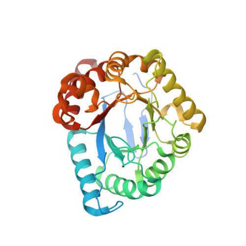
4NIR

Crystal structure of B. anthracis DHPS with compound 6: 3-[6-(trifluoromethyl)-1H-benzimidazol-2-yl]propan-1-ol


Experimental Data Snapshot

  • Method: X-RAY DIFFRACTION
  • Resolution: 1.77 Å
  • R-Value Free: 0.220 
  • R-Value Work: 0.192 
  • R-Value Observed: 0.194 

Starting Model: experimental
View more details

wwPDB Validation   3D Report Full Report


Ligand Structure Quality Assessment 


This is version 1.3 of the entry. See complete history


Literature

Identification and characterization of an allosteric inhibitory site on dihydropteroate synthase.

Hammoudeh, D.I.Date, M.Yun, M.K.Zhang, W.Boyd, V.A.Viacava Follis, A.Griffith, E.Lee, R.E.Bashford, D.White, S.W.

(2014) ACS Chem Biol 9: 1294-1302

  • DOI: https://doi.org/10.1021/cb500038g
  • Primary Citation of Related Structures:  
    4NHV, 4NIL, 4NIR, 4NL1

  • PubMed Abstract: 

    The declining effectiveness of current antibiotics due to the emergence of resistant bacterial strains dictates a pressing need for novel classes of antimicrobial therapies, preferably against molecular sites other than those in which resistance mutations have developed. Dihydropteroate synthase (DHPS) catalyzes a crucial step in the bacterial pathway of folic acid synthesis, a pathway that is absent in higher vertebrates. As the target of the sulfonamide class of drugs that were highly effective until resistance mutations arose, DHPS is known to be a valuable bacterial Achilles heel that is being further exploited for antibiotic development. Here, we report the discovery of the first known allosteric inhibitor of DHPS. NMR and crystallographic studies reveal that it engages a previously unknown binding site at the dimer interface. Kinetic data show that this inhibitor does not prevent substrate binding but rather exerts its effect at a later step in the catalytic cycle. Molecular dynamics simulations and quasi-harmonic analyses suggest that the effect of inhibitor binding is transmitted from the dimer interface to the active-site loops that are known to assume an obligatory ordered substructure during catalysis. Together with the kinetics results, these structural and dynamics data suggest an inhibitory mechanism in which binding at the dimer interface impacts loop movements that are required for product release. Our results potentially provide a novel target site for the development of new antibiotics.


  • Organizational Affiliation

    Departments of †Structural Biology and ‡Chemical Biology and Therapeutics, St. Jude Children's Research Hospital , Memphis, Tennessee 38105, United States.


Macromolecules
Find similar proteins by:  (by identity cutoff)  |  3D Structure
Entity ID: 1
MoleculeChains Sequence LengthOrganismDetailsImage
Dihydropteroate Synthase
A, B
297Bacillus anthracisMutation(s): 0 
Gene Names: folPBAS0071BA_0071GBAA_0071
EC: 2.5.1.15
UniProt
Find proteins for Q81VW8 (Bacillus anthracis)
Explore Q81VW8 
Go to UniProtKB:  Q81VW8
Entity Groups  
Sequence Clusters30% Identity50% Identity70% Identity90% Identity95% Identity100% Identity
UniProt GroupQ81VW8
Sequence Annotations
Expand
  • Reference Sequence
Small Molecules
Ligands 2 Unique
IDChains Name / Formula / InChI Key2D Diagram3D Interactions
6DH
Query on 6DH

Download Ideal Coordinates CCD File 
I [auth A],
J [auth A],
P [auth B]
3-[6-(trifluoromethyl)-1H-benzimidazol-2-yl]propan-1-ol
C11 H11 F3 N2 O
FDRMNTVCDKSRNL-UHFFFAOYSA-N
SO4
Query on SO4

Download Ideal Coordinates CCD File 
C [auth A]
D [auth A]
E [auth A]
F [auth A]
G [auth A]
C [auth A],
D [auth A],
E [auth A],
F [auth A],
G [auth A],
H [auth A],
K [auth B],
L [auth B],
M [auth B],
N [auth B],
O [auth B]
SULFATE ION
O4 S
QAOWNCQODCNURD-UHFFFAOYSA-L
Experimental Data & Validation

Experimental Data

  • Method: X-RAY DIFFRACTION
  • Resolution: 1.77 Å
  • R-Value Free: 0.220 
  • R-Value Work: 0.192 
  • R-Value Observed: 0.194 
  • Space Group: P 62 2 2
Unit Cell:
Length ( Å )Angle ( ˚ )
a = 98.338α = 90
b = 98.338β = 90
c = 263.415γ = 120
Software Package:
Software NamePurpose
DENZOdata reduction
SCALEPACKdata scaling
PHENIXrefinement
PDB_EXTRACTdata extraction
SERGUIdata collection
PHENIXphasing

Structure Validation

View Full Validation Report


Ligand Structure Quality Assessment 


Entry History 

Deposition Data

Revision History  (Full details and data files)

  • Version 1.0: 2014-05-14
    Type: Initial release
  • Version 1.1: 2014-08-06
    Changes: Database references
  • Version 1.2: 2017-11-22
    Changes: Refinement description
  • Version 1.3: 2023-09-20
    Changes: Data collection, Database references, Derived calculations, Refinement description