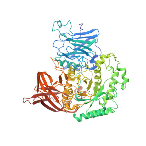
4XPQ

Crystal structure of Pedobacter saltans GH31 alpha-galactosidase complexed with L-fucose


Experimental Data Snapshot

  • Method: X-RAY DIFFRACTION
  • Resolution: 1.85 Å
  • R-Value Free: 0.168 
  • R-Value Work: 0.141 
  • R-Value Observed: 0.142 

Starting Model: experimental
View more details

wwPDB Validation   3D Report Full Report


Ligand Structure Quality Assessment 


This is version 1.4 of the entry. See complete history


Literature

Structural and biochemical characterization of novel bacterial alpha-galactosidases belonging to glycoside hydrolase family 31

Miyazaki, T.Ishizaki, Y.Ichikawa, M.Nishikawa, A.Tonozuka, T.

(2015) Biochem J 469: 145-158

  • DOI: https://doi.org/10.1042/BJ20150261
  • Primary Citation of Related Structures:  
    4XPO, 4XPP, 4XPQ, 4XPR, 4XPS

  • PubMed Abstract: 

    Glycoside hydrolase family 31 (GH31) proteins have been reportedly identified as exo-α-glycosidases with activity for α-glucosides and α-xylosides. We focused on a GH31 subfamily, which contains proteins with low sequence identity (<24%) to the previously reported GH31 glycosidases and characterized two enzymes from Pedobacter heparinus and Pedobacter saltans. The enzymes unexpectedly exhibited α-galactosidase activity, but were not active on α-glucosides and α-xylosides. The crystal structures of one of the enzymes, PsGal31A, in unliganded form and in complexes with D-galactose or L-fucose and the catalytic nucleophile mutant in unliganded form and in complex with p-nitrophenyl-α-D-galactopyranoside, were determined at 1.85-2.30 Å (1 Å=0.1 nm) resolution. The overall structure of PsGal31A contains four domains and the catalytic domain adopts a (β/α)8-barrel fold that resembles the structures of other GH31 enzymes. Two catalytic aspartic acid residues are structurally conserved in the enzymes, whereas most residues forming the active site differ from those of GH31 α-glucosidases and α-xylosidases. PsGal31A forms a dimer via a unique loop that is not conserved in other reported GH31 enzymes; this loop is involved in its aglycone specificity and in binding L-fucose. Considering potential genes for α-L-fucosidases and carbohydrate-related proteins within the vicinity of Pedobacter Gal31, the identified Gal31 enzymes are likely to function in a novel sugar degradation system. This is the first report of α-galactosidases which belong to GH31 family.


  • Organizational Affiliation

    Department of Applied Biological Science, Tokyo University of Agriculture and Technology, 3-5-8 Saiwai-cho, Fuchu, Tokyo 183-8509, Japan.


Macromolecules
Find similar proteins by:  (by identity cutoff)  |  3D Structure
Entity ID: 1
MoleculeChains Sequence LengthOrganismDetailsImage
Alpha-glucosidase720Pseudopedobacter saltansMutation(s): 0 
EC: 3.2.1.22
UniProt
Find proteins for A0A0F7R6D6 (Pseudopedobacter saltans)
Explore A0A0F7R6D6 
Go to UniProtKB:  A0A0F7R6D6
Entity Groups  
Sequence Clusters30% Identity50% Identity70% Identity90% Identity95% Identity100% Identity
UniProt GroupA0A0F7R6D6
Sequence Annotations
Expand
  • Reference Sequence
Small Molecules
Ligands 3 Unique
IDChains Name / Formula / InChI Key2D Diagram3D Interactions
FUL
Query on FUL

Download Ideal Coordinates CCD File 
C [auth A],
D [auth A],
E [auth A]
beta-L-fucopyranose
C6 H12 O5
SHZGCJCMOBCMKK-KGJVWPDLSA-N
TRS
Query on TRS

Download Ideal Coordinates CCD File 
B [auth A]2-AMINO-2-HYDROXYMETHYL-PROPANE-1,3-DIOL
C4 H12 N O3
LENZDBCJOHFCAS-UHFFFAOYSA-O
EDO
Query on EDO

Download Ideal Coordinates CCD File 
F [auth A],
G [auth A],
H [auth A],
I [auth A],
J [auth A]
1,2-ETHANEDIOL
C2 H6 O2
LYCAIKOWRPUZTN-UHFFFAOYSA-N
Experimental Data & Validation

Experimental Data

  • Method: X-RAY DIFFRACTION
  • Resolution: 1.85 Å
  • R-Value Free: 0.168 
  • R-Value Work: 0.141 
  • R-Value Observed: 0.142 
  • Space Group: P 43 21 2
Unit Cell:
Length ( Å )Angle ( ˚ )
a = 112.004α = 90
b = 112.004β = 90
c = 114.418γ = 90
Software Package:
Software NamePurpose
REFMACrefinement
HKL-2000data processing
HKL-2000data scaling
MOLREPphasing

Structure Validation

View Full Validation Report


Ligand Structure Quality Assessment 


Entry History 

Deposition Data

Revision History  (Full details and data files)

  • Version 1.0: 2015-05-20
    Type: Initial release
  • Version 1.1: 2015-09-09
    Changes: Database references
  • Version 1.2: 2020-02-05
    Changes: Data collection, Database references, Derived calculations
  • Version 1.3: 2020-07-29
    Type: Remediation
    Reason: Carbohydrate remediation
    Changes: Data collection, Derived calculations, Structure summary
  • Version 1.4: 2023-11-08
    Changes: Data collection, Database references, Refinement description, Structure summary