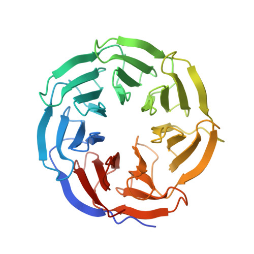
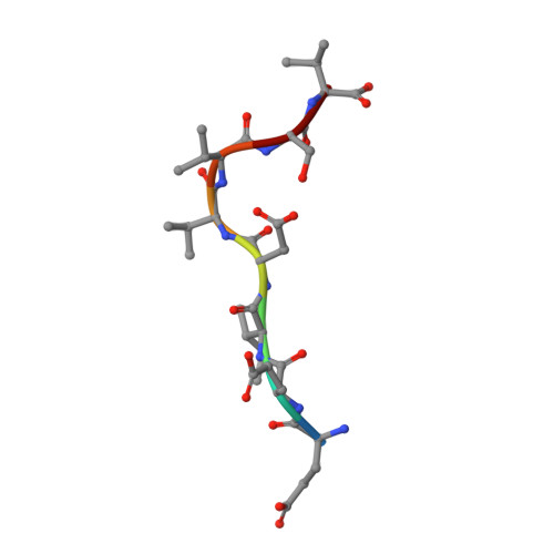
4Y7R

Crystal structure of WDR5 in complex with MYC MbIIIb peptide


Experimental Data Snapshot

  • Method: X-RAY DIFFRACTION
  • Resolution: 1.90 Å
  • R-Value Free: 0.188 
  • R-Value Work: 0.159 
  • R-Value Observed: 0.160 

Starting Model: experimental
View more details

wwPDB Validation   3D Report Full Report


This is version 1.3 of the entry. See complete history


Literature

Interaction with WDR5 Promotes Target Gene Recognition and Tumorigenesis by MYC.

Thomas, L.R.Wang, Q.Grieb, B.C.Phan, J.Foshage, A.M.Sun, Q.Olejniczak, E.T.Clark, T.Dey, S.Lorey, S.Alicie, B.Howard, G.C.Cawthon, B.Ess, K.C.Eischen, C.M.Zhao, Z.Fesik, S.W.Tansey, W.P.

(2015) Mol Cell 58: 440-452

  • DOI: https://doi.org/10.1016/j.molcel.2015.02.028
  • Primary Citation of Related Structures:  
    4Y7R

  • PubMed Abstract: 

    MYC is an oncoprotein transcription factor that is overexpressed in the majority of malignancies. The oncogenic potential of MYC stems from its ability to bind regulatory sequences in thousands of target genes, which depends on interaction of MYC with its obligate partner, MAX. Here, we show that broad association of MYC with chromatin also depends on interaction with the WD40-repeat protein WDR5. MYC binds WDR5 via an evolutionarily conserved "MYC box IIIb" motif that engages a shallow, hydrophobic cleft on the surface of WDR5. Structure-guided mutations in MYC that disrupt interaction with WDR5 attenuate binding of MYC at ∼80% of its chromosomal locations and disable its ability to promote induced pluripotent stem cell formation and drive tumorigenesis. Our data reveal WDR5 as a key determinant for MYC recruitment to chromatin and uncover a tractable target for the discovery of anticancer therapies against MYC-driven tumors.


  • Organizational Affiliation

    Department of Cell and Developmental Biology, Vanderbilt University School of Medicine, Nashville, TN 37232, USA.


Macromolecules
Find similar proteins by:  (by identity cutoff)  |  3D Structure
Entity ID: 1
MoleculeChains Sequence LengthOrganismDetailsImage
WD repeat-containing protein 5313Homo sapiensMutation(s): 0 
Gene Names: WDR5BIG3
UniProt & NIH Common Fund Data Resources
Find proteins for P61964 (Homo sapiens)
Explore P61964 
Go to UniProtKB:  P61964
PHAROS:  P61964
GTEx:  ENSG00000196363 
Entity Groups  
Sequence Clusters30% Identity50% Identity70% Identity90% Identity95% Identity100% Identity
UniProt GroupP61964
Sequence Annotations
Expand
  • Reference Sequence

Find similar proteins by:  Sequence   |   3D Structure  

Entity ID: 2
MoleculeChains Sequence LengthOrganismDetailsImage
MYC MbIIIb peptide8synthetic constructMutation(s): 0 
UniProt & NIH Common Fund Data Resources
Find proteins for P01106 (Homo sapiens)
Explore P01106 
Go to UniProtKB:  P01106
PHAROS:  P01106
GTEx:  ENSG00000136997 
Entity Groups  
UniProt GroupP01106
Sequence Annotations
Expand
  • Reference Sequence
Experimental Data & Validation

Experimental Data

  • Method: X-RAY DIFFRACTION
  • Resolution: 1.90 Å
  • R-Value Free: 0.188 
  • R-Value Work: 0.159 
  • R-Value Observed: 0.160 
  • Space Group: P 21 21 2
Unit Cell:
Length ( Å )Angle ( ˚ )
a = 80.762α = 90
b = 87.177β = 90
c = 44.914γ = 90
Software Package:
Software NamePurpose
PHENIXrefinement
HKL-2000data collection
HKL-2000data reduction
HKL-2000data scaling
PHASERphasing

Structure Validation

View Full Validation Report


Entry History & Funding Information

Deposition Data


Funding OrganizationLocationGrant Number
National Institutes of Health/Office of the DirectorUnited States5DP1OD006933

Revision History  (Full details and data files)

  • Version 1.0: 2015-04-15
    Type: Initial release
  • Version 1.1: 2015-05-20
    Changes: Database references
  • Version 1.2: 2017-09-20
    Changes: Author supporting evidence, Database references, Derived calculations, Source and taxonomy
  • Version 1.3: 2023-09-27
    Changes: Data collection, Database references, Refinement description