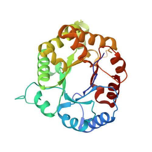
Structure-Function Studies of Hydrophobic Residues That Clamp a Basic Glutamate Side Chain during Catalysis by Triosephosphate Isomerase.
Richard, J.P., Amyes, T.L., Malabanan, M.M., Zhai, X., Kim, K.J., Reinhardt, C.J., Wierenga, R.K., Drake, E.J., Gulick, A.M.(2016) Biochemistry 55: 3036-3047
- PubMed: 27149328
- DOI: https://doi.org/10.1021/acs.biochem.6b00311
- Primary Citation of Related Structures:
5I3F, 5I3G, 5I3H, 5I3I, 5I3J, 5I3K - PubMed Abstract:
Kinetic parameters are reported for the reactions of whole substrates (kcat/Km, M(-1) s(-1)) (R)-glyceraldehyde 3-phosphate (GAP) and dihydroxyacetone phosphate (DHAP) and for the substrate pieces [(kcat/Km)E·HPi/Kd, M(-2) s(-1)] glycolaldehyde (GA) and phosphite dianion (HPi) catalyzed by the I172A/L232A mutant of triosephosphate isomerase from Trypanosoma brucei brucei (TbbTIM). A comparison with the corresponding parameters for wild-type, I172A, and L232A TbbTIM-catalyzed reactions shows that the effect of I172A and L232A mutations on ΔG(⧧) for the wild-type TbbTIM-catalyzed reactions of the substrate pieces is nearly the same as the effect of the same mutations on TbbTIM previously mutated at the second side chain. This provides strong evidence that mutation of the first hydrophobic side chain does not affect the functioning of the second side chain in catalysis of the reactions of the substrate pieces. By contrast, the effects of I172A and L232A mutations on ΔG(⧧) for wild-type TbbTIM-catalyzed reactions of the whole substrate are different from the effect of the same mutations on TbbTIM previously mutated at the second side chain. This is due to the change in the rate-determining step that determines the barrier to the isomerization reaction. X-ray crystal structures are reported for I172A, L232A, and I172A/L232A TIMs and for the complexes of these mutants to the intermediate analogue phosphoglycolate (PGA). The structures of the PGA complexes with wild-type and mutant enzymes are nearly superimposable, except that the space opened by replacement of the hydrophobic side chain is occupied by a water molecule that lies ∼3.5 Å from the basic side chain of Glu167. The new water at I172A mutant TbbTIM provides a simple rationalization for the increase in the activation barrier ΔG(⧧) observed for mutant enzyme-catalyzed reactions of the whole substrate and substrate pieces. By contrast, the new water at the L232A mutant does not predict the decrease in ΔG(⧧) observed for the mutant enzyme-catalyzed reactions of the substrate piece GA.
- Department of Chemistry, University at Buffalo, State University of New York , Buffalo, New York 14260, United States.
Organizational Affiliation:


