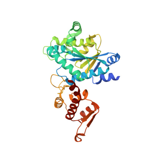
5NSF | pdb_00005nsf

Structure of AzuAla


Experimental Data Snapshot

  • Method: X-RAY DIFFRACTION
  • Resolution: 2.43 Å
  • R-Value Free: 
    0.240 (Depositor), 0.240 (DCC) 
  • R-Value Work: 
    0.182 (Depositor), 0.180 (DCC) 
  • R-Value Observed: 
    0.185 (Depositor) 

Starting Model: experimental
View more details

wwPDB Validation   3D Report Full Report


Ligand Structure Quality Assessment 

Created with Raphaël 2.3.0Worse 01 BetterLigand structure goodness of fit to experimental dataBest fitted 96ZClick on this verticalbar to view details

This is version 1.4 of the entry. See complete history


Literature

Site-Resolved Observation of Vibrational Energy Transfer Using a Genetically Encoded Ultrafast Heater.

Baumann, T.Hauf, M.Schildhauer, F.Eberl, K.B.Durkin, P.M.Deniz, E.Loffler, J.G.Acevedo-Rocha, C.G.Jaric, J.Martins, B.M.Dobbek, H.Bredenbeck, J.Budisa, N.

(2019) Angew Chem Int Ed Engl 58: 2899-2903

  • DOI: https://doi.org/10.1002/anie.201812995
  • Primary Citation of Related Structures:  
    5NSF

  • PubMed Abstract: 

    Allosteric information transfer in proteins has been linked to distinct vibrational energy transfer (VET) pathways in a number of theoretical studies. Experimental evidence for such pathways, however, is sparse because site-selective injection of vibrational energy into a protein, that is, localized heating, is required for their investigation. Here, we solved this problem by the site-specific incorporation of the non-canonical amino acid β-(1-azulenyl)-l-alanine (AzAla) through genetic code expansion. As an exception to Kasha's rule, AzAla undergoes ultrafast internal conversion and heating after S 1 excitation while upon S 2 excitation, it serves as a fluorescent label. We equipped PDZ3, a protein interaction domain of postsynaptic density protein 95, with this ultrafast heater at two distinct positions. We indeed observed VET from the incorporation sites in the protein to a bound peptide ligand on the picosecond timescale by ultrafast IR spectroscopy. This approach based on genetically encoded AzAla paves the way for detailed studies of VET and its role in a wide range of proteins.


  • Organizational Affiliation

    Institut für Chemie, Technische Universität Berlin, Müller-Breslau-Str. 10, 10623, Berlin, Germany.


Macromolecules
Find similar proteins by:  (by identity cutoff)  |  3D Structure
Entity ID: 1
MoleculeChains Sequence LengthOrganismDetailsImage
Tyrosine--tRNA ligaseA [auth B],
B [auth A],
C,
D
318Methanocaldococcus jannaschii DSM 2661Mutation(s): 0 
Gene Names: tyrSMJ0389
EC: 6.1.1.1
UniProt
Find proteins for Q57834 (Methanocaldococcus jannaschii (strain ATCC 43067 / DSM 2661 / JAL-1 / JCM 10045 / NBRC 100440))
Explore Q57834 
Go to UniProtKB:  Q57834
Entity Groups  
Sequence Clusters30% Identity50% Identity70% Identity90% Identity95% Identity100% Identity
UniProt GroupQ57834
Sequence Annotations
Expand
  • Reference Sequence
Small Molecules
Ligands 4 Unique
IDChains Name / Formula / InChI Key2D Diagram3D Interactions
96Z
Query on 96Z

Download Ideal Coordinates CCD File 
E [auth B],
H [auth A],
M [auth C],
N [auth D]
(2~{S})-2-azanyl-3-(2,6-dihydroazulen-1-yl)propanoic acid
C13 H15 N O2
HICXVJKDDWMICV-LBPRGKRZSA-N
PG4
Query on PG4

Download Ideal Coordinates CCD File 
L [auth A]TETRAETHYLENE GLYCOL
C8 H18 O5
UWHCKJMYHZGTIT-UHFFFAOYSA-N
GOL
Query on GOL

Download Ideal Coordinates CCD File 
F [auth B],
I [auth A]
GLYCEROL
C3 H8 O3
PEDCQBHIVMGVHV-UHFFFAOYSA-N
CA
Query on CA

Download Ideal Coordinates CCD File 
G [auth B],
J [auth A],
K [auth A]
CALCIUM ION
Ca
BHPQYMZQTOCNFJ-UHFFFAOYSA-N
Experimental Data & Validation

Experimental Data

  • Method: X-RAY DIFFRACTION
  • Resolution: 2.43 Å
  • R-Value Free:  0.240 (Depositor), 0.240 (DCC) 
  • R-Value Work:  0.182 (Depositor), 0.180 (DCC) 
  • R-Value Observed: 0.185 (Depositor) 
Space Group: P 1
Unit Cell:
Length ( Å )Angle ( ˚ )
a = 56.14α = 85.7
b = 70.633β = 89.75
c = 77.624γ = 83.42
Software Package:
Software NamePurpose
PHENIXrefinement
XDSdata reduction
XDSdata scaling
PHASERphasing

Structure Validation

View Full Validation Report


Ligand Structure Quality Assessment 

Created with Raphaël 2.3.0Worse 01 BetterLigand structure goodness of fit to experimental dataBest fitted 96ZClick on this verticalbar to view details

Entry History 

Deposition Data

Revision History  (Full details and data files)

  • Version 1.0: 2019-01-16
    Type: Initial release
  • Version 1.1: 2019-02-13
    Changes: Data collection, Database references
  • Version 1.2: 2019-02-27
    Changes: Data collection, Database references
  • Version 1.3: 2019-10-16
    Changes: Data collection
  • Version 1.4: 2024-01-17
    Changes: Data collection, Database references, Derived calculations, Refinement description