5TS1

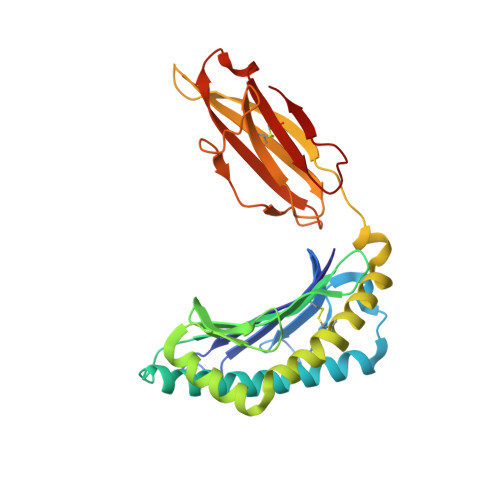
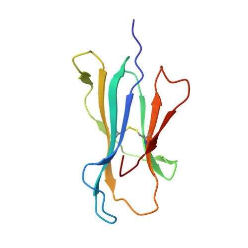
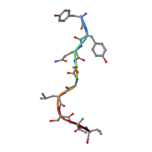
Crystal structure of MHC-I H2-KD complexed with peptides of Mycobacterial tuberculosis (YYQSGLSIV)


Experimental Data Snapshot

  • Method: X-RAY DIFFRACTION
  • Resolution: 2.30 Å
  • R-Value Free: 0.224 
  • R-Value Work: 0.187 
  • R-Value Observed: 0.189 

wwPDB Validation   3D Report Full Report


This is version 2.1 of the entry. See complete history


Literature

MHC-restricted Ag85B-specific CD8+T cells are enhanced by recombinant BCG prime and DNA boost immunization in mice.

Komine-Aizawa, S.Jiang, J.Mizuno, S.Hayakawa, S.Matsuo, K.Boyd, L.F.Margulies, D.H.Honda, M.

(2019) Eur J Immunol 

  • DOI: https://doi.org/10.1002/eji.201847988
  • Primary Citation of Related Structures:  
    5TRZ, 5TS1

  • PubMed Abstract: 

    Despite efforts to develop effective treatments and vaccines, Mycobacterium tuberculosis (Mtb), particularly pulmonary Mtb, continues to provide major health challenges worldwide. To improve immunization against the persistent health challenge of Mtb infection, we have studied the CD8 + T cell response to Bacillus Calmette-Guérin (BCG) and recombinant BCG (rBCG) in mice. Here, we generated CD8 + T cells with an rBCG-based vaccine encoding the Ag85B protein of M. kansasii, termed rBCG-Mkan85B, followed by boosting with plasmid DNA expressing the Ag85B gene (DNA-Mkan85B). We identified two MHC-I (H2-K d )-restricted epitopes that induce cross-reactive responses to Mtb and other related mycobacteria in both BALB/c (H2 d ) and CB6F1 (H2 b/d ) mice. The H2-K d -restricted peptide epitopes elicited polyfunctional CD8 + T cell responses that were also highly cross-reactive with those of other proteins of the Ag85 complex. Tetramer staining indicated that the two H2-K d -restricted epitopes elicit distinct CD8 + T cell populations, a result explained by the X-ray structure of the two peptide/H2-K d complexes. These results suggest that rBCG-Mkan85B vector-based immunization and DNA-Mkan85B boost may enhance CD8 + T cell response to Mtb, and might help to overcome the limited effectiveness of the current BCG in eliciting tuberculosis immunity.


  • Organizational Affiliation

    Division of Microbiology, Department of Pathology and Microbiology, Nihon University School of Medicine, Nihon University, Tokyo, Japan.


Macromolecules
Find similar proteins by:  (by identity cutoff)  |  3D Structure
Entity ID: 1
MoleculeChains Sequence LengthOrganismDetailsImage
H-2 class I histocompatibility antigen, K-D alpha chainA,
D [auth C],
G [auth E],
J [auth G]
275Mus musculusMutation(s): 0 
Gene Names: H2-K1H2-K
UniProt
Find proteins for P01902 (Mus musculus)
Explore P01902 
Go to UniProtKB:  P01902
Entity Groups  
Sequence Clusters30% Identity50% Identity70% Identity90% Identity95% Identity100% Identity
UniProt GroupP01902
Sequence Annotations
Expand
  • Reference Sequence
Find similar proteins by:  (by identity cutoff)  |  3D Structure
Entity ID: 2
MoleculeChains Sequence LengthOrganismDetailsImage
Beta-2-microglobulinB,
E [auth D],
H [auth F],
K [auth H]
100Homo sapiensMutation(s): 0 
Gene Names: B2MCDABP0092HDCMA22P
UniProt & NIH Common Fund Data Resources
Find proteins for P61769 (Homo sapiens)
Explore P61769 
Go to UniProtKB:  P61769
PHAROS:  P61769
GTEx:  ENSG00000166710 
Entity Groups  
Sequence Clusters30% Identity50% Identity70% Identity90% Identity95% Identity100% Identity
UniProt GroupP61769
Sequence Annotations
Expand
  • Reference Sequence

Find similar proteins by:  Sequence   |   3D Structure  

Entity ID: 3
MoleculeChains Sequence LengthOrganismDetailsImage
Peptide (P9) of Mtb85B (Mycobacterium tuberculosis) YYQSGLSIVC [auth P],
F [auth Q],
I [auth R],
L [auth S]
9Mycobacterium tuberculosis H37RvMutation(s): 0 
EC: 2.3.1.20 (UniProt), 2.3.1.122 (UniProt)
UniProt
Find proteins for P9WQP1 (Mycobacterium tuberculosis (strain ATCC 25618 / H37Rv))
Explore P9WQP1 
Go to UniProtKB:  P9WQP1
Entity Groups  
UniProt GroupP9WQP1
Sequence Annotations
Expand
  • Reference Sequence
Small Molecules
Ligands 2 Unique
IDChains Name / Formula / InChI Key2D Diagram3D Interactions
GOL
Query on GOL

Download Ideal Coordinates CCD File 
M [auth A]
N [auth A]
R [auth D]
S [auth F]
T [auth G]
M [auth A],
N [auth A],
R [auth D],
S [auth F],
T [auth G],
U [auth H]
GLYCEROL
C3 H8 O3
PEDCQBHIVMGVHV-UHFFFAOYSA-N
EDO
Query on EDO

Download Ideal Coordinates CCD File 
O [auth A],
P [auth C],
Q [auth C]
1,2-ETHANEDIOL
C2 H6 O2
LYCAIKOWRPUZTN-UHFFFAOYSA-N
Experimental Data & Validation

Experimental Data

  • Method: X-RAY DIFFRACTION
  • Resolution: 2.30 Å
  • R-Value Free: 0.224 
  • R-Value Work: 0.187 
  • R-Value Observed: 0.189 
  • Space Group: P 1
Unit Cell:
Length ( Å )Angle ( ˚ )
a = 47.302α = 89.97
b = 88.96β = 93.83
c = 109.947γ = 90.04
Software Package:
Software NamePurpose
PHENIXrefinement
XDSdata reduction
XDSdata scaling
PHASERphasing

Structure Validation

View Full Validation Report


Entry History 

Deposition Data

Revision History  (Full details and data files)

  • Version 1.0: 2018-05-09
    Type: Initial release
  • Version 1.1: 2019-08-14
    Changes: Data collection, Database references
  • Version 2.0: 2020-02-05
    Changes: Atomic model, Data collection, Derived calculations, Refinement description
  • Version 2.1: 2024-11-06
    Changes: Data collection, Database references, Structure summary