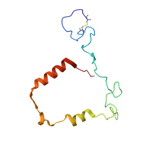
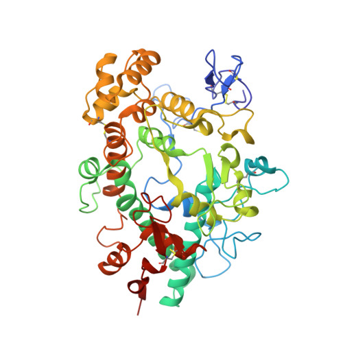
5WDJ

CRYSTAL STRUCTURE OF MYELOPEROXIDASE SUBFORM C (MPO) COMPLEX WITH COMPOUND-6 AKA 7-(BENZYLOXY)-1H-[1,2, 3]TRIAZOLO[4,5-D]PYRIMIDIN-5-AMINE


Experimental Data Snapshot

  • Method: X-RAY DIFFRACTION
  • Resolution: 2.40 Å
  • R-Value Free: 0.248 
  • R-Value Work: 0.194 
  • R-Value Observed: 0.197 

wwPDB Validation   3D Report Full Report


Ligand Structure Quality Assessment 


This is version 2.1 of the entry. See complete history


Literature

Triazolopyrimidines identified as reversible myeloperoxidase inhibitors.

Duclos, F.Abell, L.M.Harden, D.G.Pike, K.Nowak, K.Locke, G.A.Duke, G.J.Liu, X.Fernando, G.Shaw, S.A.Vokits, B.P.Wurtz, N.R.Viet, A.Valente, M.N.Stachura, S.Sleph, P.Khan, J.A.Gao, J.Dongre, A.R.Zhao, L.Wexler, R.R.Gordon, D.A.Kick, E.K.

(2017) Medchemcomm 8: 2093-2099

  • DOI: https://doi.org/10.1039/c7md00268h
  • Primary Citation of Related Structures:  
    5WDJ

  • PubMed Abstract: 

    Myeloperoxidase, a mammalian peroxidase involved in the immune system as an anti-microbial first responder, can produce hypochlorous acid in response to invading pathogens. Myeloperoxidase has been implicated in several chronic pathological diseases due to the chronic production of hypochlorous acid, as well as other reactive radical species. A high throughput screen and triaging protocol was developed to identify a reversible inhibitor of myeloperoxidase toward the potential treatment of chronic diseases such as atherosclerosis. The identification and characterization of a reversible myeloperoxidase inhibitor, 7-(benzyloxy)-3 H -[1,2,3]triazolo[4,5- d ]pyrimidin-5-amine is described.


  • Organizational Affiliation

    Bristol-Myers Squibb Company , P.O. Box 5400 , Princeton , New Jersey 08543-5400 , USA . Email: Franck.Duclos@Bms.com.


Macromolecules
Find similar proteins by:  (by identity cutoff)  |  3D Structure
Entity ID: 1
MoleculeChains Sequence LengthOrganismDetailsImage
MyeloperoxidaseA,
C [auth D]
105Homo sapiensMutation(s): 0 
Gene Names: MPO
EC: 1.11.2.2
UniProt & NIH Common Fund Data Resources
Find proteins for P05164 (Homo sapiens)
Explore P05164 
Go to UniProtKB:  P05164
PHAROS:  P05164
GTEx:  ENSG00000005381 
Entity Groups  
Sequence Clusters30% Identity50% Identity70% Identity90% Identity95% Identity100% Identity
UniProt GroupP05164
Sequence Annotations
Expand
  • Reference Sequence
Find similar proteins by:  (by identity cutoff)  |  3D Structure
Entity ID: 2
MoleculeChains Sequence LengthOrganismDetailsImage
MyeloperoxidaseB,
D [auth E]
467Homo sapiensMutation(s): 0 
Gene Names: MPO
EC: 1.11.2.2
UniProt & NIH Common Fund Data Resources
Find proteins for P05164 (Homo sapiens)
Explore P05164 
Go to UniProtKB:  P05164
PHAROS:  P05164
GTEx:  ENSG00000005381 
Entity Groups  
Sequence Clusters30% Identity50% Identity70% Identity90% Identity95% Identity100% Identity
UniProt GroupP05164
Glycosylation
Glycosylation Sites: 3Go to GlyGen: P05164-1
Sequence Annotations
Expand
  • Reference Sequence
Oligosaccharides

Help

Entity ID: 3
MoleculeChains Length2D Diagram Glycosylation3D Interactions
alpha-D-mannopyranose-(1-3)-beta-D-mannopyranose-(1-4)-2-acetamido-2-deoxy-beta-D-glucopyranoseE [auth C]3N/A
Glycosylation Resources
GlyTouCan:  G29301NP
GlyCosmos:  G29301NP
GlyGen:  G29301NP
Entity ID: 4
MoleculeChains Length2D Diagram Glycosylation3D Interactions
beta-D-mannopyranose-(1-4)-2-acetamido-2-deoxy-beta-D-glucopyranose-(1-4)-2-acetamido-2-deoxy-beta-D-glucopyranose
F
3N-Glycosylation
Glycosylation Resources
GlyTouCan:  G15407YE
GlyCosmos:  G15407YE
GlyGen:  G15407YE
Small Molecules
Ligands 7 Unique
IDChains Name / Formula / InChI Key2D Diagram3D Interactions
HEM
Query on HEM

Download Ideal Coordinates CCD File 
I [auth A],
R [auth D]
PROTOPORPHYRIN IX CONTAINING FE
C34 H32 Fe N4 O4
KABFMIBPWCXCRK-RGGAHWMASA-L
AEY
Query on AEY

Download Ideal Coordinates CCD File 
G [auth A],
Y [auth E]
7-(benzyloxy)-1H-[1,2,3]triazolo[4,5-d]pyrimidin-5-amine
C11 H10 N6 O
ZHTBAYPUCDCQPX-UHFFFAOYSA-N
NAG
Query on NAG

Download Ideal Coordinates CCD File 
K [auth B],
M [auth B],
N [auth B],
W [auth E],
X [auth E]
2-acetamido-2-deoxy-beta-D-glucopyranose
C8 H15 N O6
OVRNDRQMDRJTHS-FMDGEEDCSA-N
MAN
Query on MAN

Download Ideal Coordinates CCD File 
O [auth B],
P [auth B],
U [auth E]
alpha-D-mannopyranose
C6 H12 O6
WQZGKKKJIJFFOK-PQMKYFCFSA-N
FUC
Query on FUC

Download Ideal Coordinates CCD File 
L [auth B],
V [auth E]
alpha-L-fucopyranose
C6 H12 O5
SHZGCJCMOBCMKK-SXUWKVJYSA-N
CA
Query on CA

Download Ideal Coordinates CCD File 
J [auth A],
T [auth D]
CALCIUM ION
Ca
BHPQYMZQTOCNFJ-UHFFFAOYSA-N
CL
Query on CL

Download Ideal Coordinates CCD File 
H [auth A],
Q [auth B],
S [auth D],
Z [auth E]
CHLORIDE ION
Cl
VEXZGXHMUGYJMC-UHFFFAOYSA-M
Modified Residues  1 Unique
IDChains TypeFormula2D DiagramParent
CSO
Query on CSO
B,
D [auth E]
L-PEPTIDE LINKINGC3 H7 N O3 SCYS
Binding Affinity Annotations 
IDSourceBinding Affinity
AEY BindingDB:  5WDJ IC50: min: 103, max: 110 (nM) from 3 assay(s)
Experimental Data & Validation

Experimental Data

  • Method: X-RAY DIFFRACTION
  • Resolution: 2.40 Å
  • R-Value Free: 0.248 
  • R-Value Work: 0.194 
  • R-Value Observed: 0.197 
  • Space Group: P 43 21 2
Unit Cell:
Length ( Å )Angle ( ˚ )
a = 103.925α = 90
b = 103.925β = 90
c = 242.276γ = 90
Software Package:
Software NamePurpose
BUSTERrefinement
PDB_EXTRACTdata extraction
HKL-2000data reduction
SCALEPACKdata scaling
PHASERphasing

Structure Validation

View Full Validation Report


Ligand Structure Quality Assessment 


Entry History 

Deposition Data

  • Released Date: 2018-04-18 
  • Deposition Author(s): Khan, J.A.

Revision History  (Full details and data files)

  • Version 1.0: 2018-04-18
    Type: Initial release
  • Version 1.1: 2018-10-31
    Changes: Data collection, Database references
  • Version 2.0: 2020-07-29
    Type: Remediation
    Reason: Carbohydrate remediation
    Changes: Advisory, Atomic model, Data collection, Derived calculations, Structure summary
  • Version 2.1: 2024-10-23
    Changes: Data collection, Database references, Derived calculations, Refinement description, Structure summary