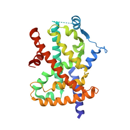
6DHA

Crystal Structure of Human PPARgamma Ligand Binding Domain in Complex with Hydroxy Pioglitazone (M-IV)


Experimental Data Snapshot

  • Method: X-RAY DIFFRACTION
  • Resolution: 1.88 Å
  • R-Value Free: 0.255 
  • R-Value Work: 0.220 
  • R-Value Observed: 0.222 

Starting Model: experimental
View more details

wwPDB Validation   3D Report Full Report


Ligand Structure Quality Assessment 


This is version 1.2 of the entry. See complete history


Literature

Structural Basis of Altered Potency and Efficacy Displayed by a Major in Vivo Metabolite of the Antidiabetic PPAR gamma Drug Pioglitazone.

Mosure, S.A.Shang, J.Eberhardt, J.Brust, R.Zheng, J.Griffin, P.R.Forli, S.Kojetin, D.J.

(2019) J Med Chem 62: 2008-2023

  • DOI: https://doi.org/10.1021/acs.jmedchem.8b01573
  • Primary Citation of Related Structures:  
    6DHA

  • PubMed Abstract: 

    Pioglitazone (Pio) is a Food and Drug Administration-approved drug for type-2 diabetes that binds and activates the nuclear receptor peroxisome proliferator-activated receptor γ (PPARγ), yet it remains unclear how in vivo Pio metabolites affect PPARγ structure and function. Here, we present a structure-function comparison of Pio and its most abundant in vivo metabolite, 1-hydroxypioglitazone (PioOH). PioOH displayed a lower binding affinity and reduced potency in co-regulator recruitment assays. X-ray crystallography and molecular docking analysis of PioOH-bound PPARγ ligand-binding domain revealed an altered hydrogen bonding network, including the formation of water-mediated bonds, which could underlie its altered biochemical phenotype. NMR spectroscopy and hydrogen/deuterium exchange mass spectrometry analysis coupled to activity assays revealed that PioOH better stabilizes the PPARγ activation function-2 (AF-2) co-activator binding surface and better enhances co-activator binding, affording slightly better transcriptional efficacy. These results indicating that Pio hydroxylation affects its potency and efficacy as a PPARγ agonist contributes to our understanding of PPARγ-drug metabolite interactions.


  • Organizational Affiliation

    Department of Integrative Structural and Computational Biology , The Scripps Research Institute , La Jolla , California 92037 , United States.


Macromolecules
Find similar proteins by:  (by identity cutoff)  |  3D Structure
Entity ID: 1
MoleculeChains Sequence LengthOrganismDetailsImage
Peroxisome proliferator-activated receptor gamma
A, B
271Homo sapiensMutation(s): 0 
Gene Names: PPARGNR1C3
UniProt & NIH Common Fund Data Resources
Find proteins for P37231 (Homo sapiens)
Explore P37231 
Go to UniProtKB:  P37231
PHAROS:  P37231
GTEx:  ENSG00000132170 
Entity Groups  
Sequence Clusters30% Identity50% Identity70% Identity90% Identity95% Identity100% Identity
UniProt GroupP37231
Sequence Annotations
Expand
  • Reference Sequence
Small Molecules
Binding Affinity Annotations 
IDSourceBinding Affinity
GFV BindingDB:  6DHA Ki: 1200 (nM) from 1 assay(s)
Kd: 708 (nM) from 1 assay(s)
IC50: 1.30e+4 (nM) from 1 assay(s)
EC50: min: 680, max: 2100 (nM) from 3 assay(s)
Experimental Data & Validation

Experimental Data

  • Method: X-RAY DIFFRACTION
  • Resolution: 1.88 Å
  • R-Value Free: 0.255 
  • R-Value Work: 0.220 
  • R-Value Observed: 0.222 
  • Space Group: C 1 2 1
Unit Cell:
Length ( Å )Angle ( ˚ )
a = 93.08α = 90
b = 61.85β = 102.73
c = 119.67γ = 90
Software Package:
Software NamePurpose
PHENIXrefinement
SCALAdata scaling
PHASERphasing
PDB_EXTRACTdata extraction
MOSFLMdata reduction

Structure Validation

View Full Validation Report


Ligand Structure Quality Assessment 


Entry History & Funding Information

Deposition Data


Funding OrganizationLocationGrant Number
National Institutes of Health/National Institute of Diabetes and Digestive and Kidney Disease (NIH/NIDDK)United StatesR01DK101871

Revision History  (Full details and data files)

  • Version 1.0: 2019-03-13
    Type: Initial release
  • Version 1.1: 2019-12-25
    Changes: Author supporting evidence
  • Version 1.2: 2023-10-11
    Changes: Data collection, Database references, Refinement description