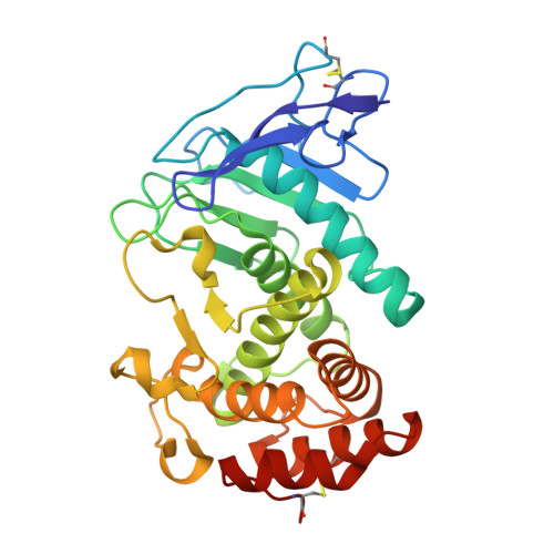
6FZX

LasB, hydroxymate Inhibitor Complex


Experimental Data Snapshot

  • Method: X-RAY DIFFRACTION
  • Resolution: 2.10 Å
  • R-Value Free: 0.213 
  • R-Value Work: 0.179 
  • R-Value Observed: 0.181 

Starting Model: experimental
View more details

wwPDB Validation   3D Report Full Report


Ligand Structure Quality Assessment 


This is version 1.3 of the entry. See complete history


Literature

Tackling Pseudomonas aeruginosa Virulence by a Hydroxamic Acid-Based LasB Inhibitor.

Kany, A.M.Sikandar, A.Yahiaoui, S.Haupenthal, J.Walter, I.Empting, M.Kohnke, J.Hartmann, R.W.

(2018) ACS Chem Biol 13: 2449-2455

  • DOI: https://doi.org/10.1021/acschembio.8b00257
  • Primary Citation of Related Structures:  
    6FZX

  • PubMed Abstract: 

    In search of novel antibiotics to combat the challenging spread of resistant pathogens, bacterial proteases represent promising targets for pathoblocker development. A common motif for protease inhibitors is the hydroxamic acid function, yet this group has often been related to unspecific inhibition of various metalloproteases. In this work, the inhibition of LasB, a harmful zinc metalloprotease secreted by Pseudomonas aeruginosa, through a hydroxamate derivative is described. The present inhibitor was developed based on a recently reported, highly selective thiol scaffold. Using X-ray crystallography, the lack of inhibition of a range of human matrix metalloproteases could be attributed to a distinct binding mode sparing the S1' pocket. The inhibitor was shown to restore the effect of the antimicrobial peptide LL-37, decrease the formation of P. aeruginosa biofilm and, for the first time for a LasB inhibitor, reduce the release of extracellular DNA. Hence, it is capable of disrupting several important bacterial resistance mechanisms. These results highlight the potential of protease inhibitors to fight bacterial infections and point out the possibility to achieve selective inhibition even with a strong zinc anchor.


  • Organizational Affiliation

    Department of Drug Design and Optimization , Helmholtz Institute for Pharmaceutical Research Saarland (HIPS) , Campus E8.1 , 66123 Saarbrücken , Germany.


Macromolecules
Find similar proteins by:  (by identity cutoff)  |  3D Structure
Entity ID: 1
MoleculeChains Sequence LengthOrganismDetailsImage
Keratinase KP2301Pseudomonas aeruginosaMutation(s): 0 
EC: 3.4.24.26
UniProt
Find proteins for P14756 (Pseudomonas aeruginosa (strain ATCC 15692 / DSM 22644 / CIP 104116 / JCM 14847 / LMG 12228 / 1C / PRS 101 / PAO1))
Explore P14756 
Go to UniProtKB:  P14756
Entity Groups  
Sequence Clusters30% Identity50% Identity70% Identity90% Identity95% Identity100% Identity
UniProt GroupP14756
Sequence Annotations
Expand
  • Reference Sequence
Small Molecules
Ligands 5 Unique
IDChains Name / Formula / InChI Key2D Diagram3D Interactions
EEK
Query on EEK

Download Ideal Coordinates CCD File 
B [auth A]~{N}-(3,4-dichlorophenyl)-~{N}'-oxidanyl-propanediamide
C9 H8 Cl2 N2 O3
ZGACHNBRFOETSO-UHFFFAOYSA-N
SO4
Query on SO4

Download Ideal Coordinates CCD File 
E [auth A]SULFATE ION
O4 S
QAOWNCQODCNURD-UHFFFAOYSA-L
GOL
Query on GOL

Download Ideal Coordinates CCD File 
F [auth A]GLYCEROL
C3 H8 O3
PEDCQBHIVMGVHV-UHFFFAOYSA-N
ZN
Query on ZN

Download Ideal Coordinates CCD File 
C [auth A]ZINC ION
Zn
PTFCDOFLOPIGGS-UHFFFAOYSA-N
CA
Query on CA

Download Ideal Coordinates CCD File 
D [auth A]CALCIUM ION
Ca
BHPQYMZQTOCNFJ-UHFFFAOYSA-N
Experimental Data & Validation

Experimental Data

  • Method: X-RAY DIFFRACTION
  • Resolution: 2.10 Å
  • R-Value Free: 0.213 
  • R-Value Work: 0.179 
  • R-Value Observed: 0.181 
  • Space Group: P 21 21 21
Unit Cell:
Length ( Å )Angle ( ˚ )
a = 43.727α = 90
b = 52.982β = 90
c = 122.519γ = 90
Software Package:
Software NamePurpose
PHENIXrefinement
XDSdata reduction
SCALAdata scaling
PHASERphasing

Structure Validation

View Full Validation Report


Ligand Structure Quality Assessment 


Entry History & Funding Information

Deposition Data


Funding OrganizationLocationGrant Number
German Research FoundationGermanyKO4116_3_1

Revision History  (Full details and data files)

  • Version 1.0: 2018-09-05
    Type: Initial release
  • Version 1.1: 2018-10-03
    Changes: Data collection, Database references
  • Version 1.2: 2024-01-17
    Changes: Data collection, Database references, Refinement description
  • Version 1.3: 2024-11-13
    Changes: Structure summary