6N6K

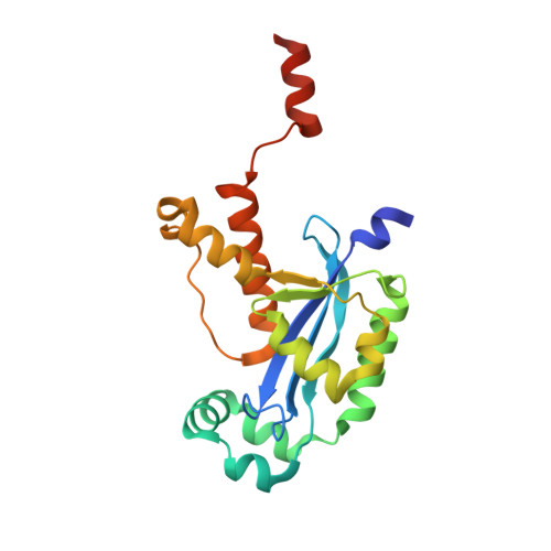
Human REXO2 bound to pAG


Experimental Data Snapshot

  • Method: X-RAY DIFFRACTION
  • Resolution: 1.42 Å
  • R-Value Free: 0.172 
  • R-Value Work: 0.153 
  • R-Value Observed: 0.154 

Starting Model: experimental
View more details

wwPDB Validation   3D Report Full Report


This is version 1.3 of the entry. See complete history


Literature

A dedicated diribonucleotidase resolves a key bottleneck for the terminal step of RNA degradation.

Kim, S.K.Lormand, J.D.Weiss, C.A.Eger, K.A.Turdiev, H.Turdiev, A.Winkler, W.C.Sondermann, H.Lee, V.T.

(2019) Elife 8

  • DOI: https://doi.org/10.7554/eLife.46313
  • Primary Citation of Related Structures:  
    6N6A, 6N6C, 6N6D, 6N6E, 6N6F, 6N6G, 6N6H, 6N6I, 6N6J, 6N6K

  • PubMed Abstract: 

    Degradation of RNA polymers, an ubiquitous process in all cells, is catalyzed by specific subsets of endo- and exoribonucleases that together recycle RNA fragments into nucleotide monophosphate. In γ-proteobacteria, 3-'5' exoribonucleases comprise up to eight distinct enzymes. Among them, Oligoribonuclease (Orn) is unique as its activity is required for clearing short RNA fragments, which is important for cellular fitness. However, the molecular basis of Orn's unique cellular function remained unclear. Here, we show that Orn exhibits exquisite substrate preference for diribonucleotides. Crystal structures of substrate-bound Orn reveal an active site optimized for diribonucleotides. While other cellular RNases process oligoribonucleotides down to diribonucleotide entities, Orn is the one and only diribonucleotidase that completes the terminal step of RNA degradation. Together, our studies indicate RNA degradation as a step-wise process with a dedicated enzyme for the clearance of a specific intermediate pool, diribonucleotides, that affects cellular physiology and viability.


  • Organizational Affiliation

    Department of Cell Biology and Molecular Genetics, University of Maryland, College Park, United States.


Macromolecules

Find similar proteins by:  (by identity cutoff)  |  3D Structure
Entity ID: 1
MoleculeChains Sequence LengthOrganismDetailsImage
RNA exonuclease 2 homolog,Small fragment nucleaseA,
C [auth B]
205Homo sapiensMutation(s): 0 
Gene Names: REXO2SFNSMFNCGI-114
EC: 3.1 (PDB Primary Data), 3.1.15 (UniProt)
UniProt & NIH Common Fund Data Resources
Find proteins for Q9Y3B8 (Homo sapiens)
Explore Q9Y3B8 
Go to UniProtKB:  Q9Y3B8
PHAROS:  Q9Y3B8
GTEx:  ENSG00000076043 
Entity Groups  
Sequence Clusters30% Identity50% Identity70% Identity90% Identity95% Identity100% Identity
UniProt GroupQ9Y3B8
Sequence Annotations
Expand
  • Reference Sequence

Find similar nucleic acids by:  Sequence   |   3D Structure  

Entity ID: 2
MoleculeChains LengthOrganismImage
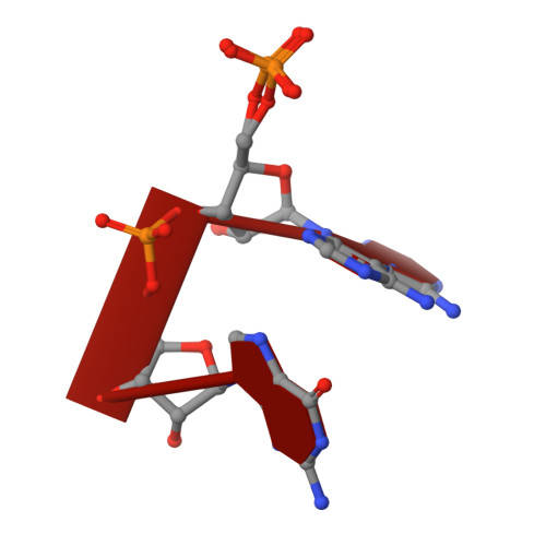
RNA (5'-R(P*AP*G)-3')B [auth D],
D [auth C]
2synthetic construct
Sequence Annotations
Expand
  • Reference Sequence
Experimental Data & Validation

Experimental Data

  • Method: X-RAY DIFFRACTION
  • Resolution: 1.42 Å
  • R-Value Free: 0.172 
  • R-Value Work: 0.153 
  • R-Value Observed: 0.154 
  • Space Group: P 21 21 21
Unit Cell:
Length ( Å )Angle ( ˚ )
a = 45.806α = 90
b = 87.423β = 90
c = 124.276γ = 90
Software Package:
Software NamePurpose
XDSdata reduction
SCALAdata scaling
PHENIXrefinement
PDB_EXTRACTdata extraction
PHENIXphasing

Structure Validation

View Full Validation Report


Entry History & Funding Information

Deposition Data


Funding OrganizationLocationGrant Number
National Institutes of Health/National Institute of General Medical Sciences (NIH/NIGMS)United StatesR01GM123609

Revision History  (Full details and data files)

  • Version 1.0: 2019-06-12
    Type: Initial release
  • Version 1.1: 2019-10-16
    Changes: Data collection, Database references
  • Version 1.2: 2020-01-01
    Changes: Author supporting evidence
  • Version 1.3: 2023-10-11
    Changes: Data collection, Database references, Derived calculations, Refinement description