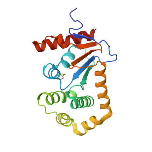
6PML

Crystal Structure of EcDsbA in complex benzyl ether 23


Experimental Data Snapshot

  • Method: X-RAY DIFFRACTION
  • Resolution: 2.00 Å
  • R-Value Free: 0.221 
  • R-Value Work: 0.182 
  • R-Value Observed: 0.185 

Starting Model: experimental
View more details

wwPDB Validation   3D Report Full Report


Ligand Structure Quality Assessment 


This is version 1.3 of the entry. See complete history


Literature

The Fragment-Based Development of a Benzofuran Hit as a New Class of Escherichia coli DsbA Inhibitors.

Duncan, L.F.Wang, G.Ilyichova, O.V.Scanlon, M.J.Heras, B.Abbott, B.M.

(2019) Molecules 24

  • DOI: https://doi.org/10.3390/molecules24203756
  • Primary Citation of Related Structures:  
    6PMF, 6PML, 6POH, 6POI, 6POQ, 6PVY, 6PVZ

  • PubMed Abstract: 

    A fragment-based drug discovery approach was taken to target the thiol-disulfide oxidoreductase enzyme DsbA from Escherichia coli ( Ec DsbA). This enzyme is critical for the correct folding of virulence factors in many pathogenic Gram-negative bacteria, and small molecule inhibitors can potentially be developed as anti-virulence compounds. Biophysical screening of a library of fragments identified several classes of fragments with affinity to Ec DsbA. One hit with high mM affinity, 2-(6-bromobenzofuran-3-yl)acetic acid ( 6 ), was chemically elaborated at several positions around the scaffold. X-ray crystal structures of the elaborated analogues showed binding in the hydrophobic binding groove adjacent to the catalytic disulfide bond of Ec DsbA. Binding affinity was calculated based on NMR studies and compounds 25 and 28 were identified as the highest affinity binders with dissociation constants ( K D ) of 326 ± 25 and 341 ± 57 µM respectively. This work suggests the potential to develop benzofuran fragments into a novel class of Ec DsbA inhibitors.


  • Organizational Affiliation

    Department of Chemistry and Physics, La Trobe Institute for Molecular Science, La Trobe University, Melbourne 3086, Australia. dunclf01@gmail.com.


Macromolecules
Find similar proteins by:  (by identity cutoff)  |  3D Structure
Entity ID: 1
MoleculeChains Sequence LengthOrganismDetailsImage
Thiol:disulfide interchange protein DsbA
A, B
189Escherichia coli K-12Mutation(s): 0 
Gene Names: dsbAdsfppfAb3860JW3832
UniProt
Find proteins for P0AEG4 (Escherichia coli (strain K12))
Explore P0AEG4 
Go to UniProtKB:  P0AEG4
Entity Groups  
Sequence Clusters30% Identity50% Identity70% Identity90% Identity95% Identity100% Identity
UniProt GroupP0AEG4
Sequence Annotations
Expand
  • Reference Sequence
Experimental Data & Validation

Experimental Data

  • Method: X-RAY DIFFRACTION
  • Resolution: 2.00 Å
  • R-Value Free: 0.221 
  • R-Value Work: 0.182 
  • R-Value Observed: 0.185 
  • Space Group: C 1 2 1
Unit Cell:
Length ( Å )Angle ( ˚ )
a = 117.54α = 90
b = 63.74β = 140.85
c = 96.19γ = 90
Software Package:
Software NamePurpose
PHENIXrefinement
MOSFLMdata reduction
SCALAdata scaling
PHASERphasing

Structure Validation

View Full Validation Report


Ligand Structure Quality Assessment 


Entry History 

Deposition Data

Revision History  (Full details and data files)

  • Version 1.0: 2019-11-06
    Type: Initial release
  • Version 1.1: 2020-10-07
    Changes: Database references
  • Version 1.2: 2023-10-11
    Changes: Data collection, Database references, Refinement description
  • Version 1.3: 2024-10-23
    Changes: Structure summary