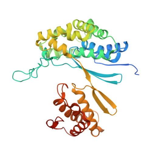
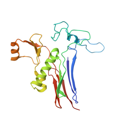
7D9X

Highly active mutant W525D of Gamma-glutamyltranspeptidase from Pseudomonas nitroreducens


Experimental Data Snapshot

  • Method: X-RAY DIFFRACTION
  • Resolution: 1.74 Å
  • R-Value Free: 0.187 
  • R-Value Work: 0.167 
  • R-Value Observed: 0.167 

Starting Model: experimental
View more details

wwPDB Validation   3D Report Full Report


Ligand Structure Quality Assessment 


This is version 1.1 of the entry. See complete history


Literature

Mutagenesis and structure-based analysis of the role of Tryptophan525 of gamma-glutamyltranspeptidase from Pseudomonas nitroreducens.

Sano, C.Itoh, T.Phumsombat, P.Hayashi, J.Wakayama, M.Hibi, T.

(2021) Biochem Biophys Res Commun 534: 286-291

  • DOI: https://doi.org/10.1016/j.bbrc.2020.11.093
  • Primary Citation of Related Structures:  
    7D9E, 7D9W, 7D9X

  • PubMed Abstract: 

    γ-Glutamyltranspeptidase (GGT) is a ubiquitous enzyme that catalyzes the hydrolysis of the γ-glutamyl linkage of γ-glutamyl compounds and the transfer of their γ-glutamyl moiety to acceptor substrates. Pseudomonas nitroreducens GGT (PnGGT) is used for the industrial synthesis of theanine, thus it is important to determine the structural basis of hydrolysis and transfer reactions and identify the acceptor site of PnGGT to improve the efficient of theanine synthesis. Our previous structural studies of PnGGT have revealed that crucial interactions between three amino acid residues, Trp385, Phe417, and Trp525, distinguish PnGGT from other GGTs. Here we report the role of Trp525 in PnGGT based on site-directed mutagenesis and structural analyses. Seven mutant variants of Trp525 were produced (W525F, W525V, W525A, W525G, W525S, W525D, and W525K), with substitution of Trp525 by nonaromatic residues resulting in dramatically reduced hydrolysis activity. All Trp525 mutants exhibited significantly increased transfer activity toward hydroxylamine with hardly any effect on acceptor substrate preference. The crystal structure of PnGGT in complex with the glutamine antagonist, 6-diazo-5-oxo-l-norleucine, revealed that Trp525 is a key residue limiting the movement of water molecules within the PnGGT active site.


  • Organizational Affiliation

    Department of Biotechnology, College of Life Sciences, Ritsumeikan University, Kusatsu, Shiga, 525-8577, Japan.


Macromolecules
Find similar proteins by:  (by identity cutoff)  |  3D Structure
Entity ID: 1
MoleculeChains Sequence LengthOrganismDetailsImage
Gamma-glutamyltransferase 1 Threonine peptidase. MEROPS family T03
A, C
371Pseudomonas nitroreducensMutation(s): 0 
Gene Names: SAMN05216209_3923
EC: 2.3.2.2 (UniProt), 3.4.19.13 (UniProt)
UniProt
Find proteins for A0A6G6IRK3 (Pseudomonas nitroreducens)
Explore A0A6G6IRK3 
Go to UniProtKB:  A0A6G6IRK3
Entity Groups  
Sequence Clusters30% Identity50% Identity70% Identity90% Identity95% Identity100% Identity
UniProt GroupA0A6G6IRK3
Sequence Annotations
Expand
  • Reference Sequence
Find similar proteins by:  (by identity cutoff)  |  3D Structure
Entity ID: 2
MoleculeChains Sequence LengthOrganismDetailsImage
Gamma-glutamyltransferase 1 Threonine peptidase. MEROPS family T03
B, D
194Pseudomonas nitroreducensMutation(s): 1 
Gene Names: SAMN05216209_3923
EC: 2.3.2.2 (UniProt), 3.4.19.13 (UniProt)
UniProt
Find proteins for A0A6G6IRK3 (Pseudomonas nitroreducens)
Explore A0A6G6IRK3 
Go to UniProtKB:  A0A6G6IRK3
Entity Groups  
Sequence Clusters30% Identity50% Identity70% Identity90% Identity95% Identity100% Identity
UniProt GroupA0A6G6IRK3
Sequence Annotations
Expand
  • Reference Sequence
Small Molecules
Ligands 3 Unique
IDChains Name / Formula / InChI Key2D Diagram3D Interactions
GOL
Query on GOL

Download Ideal Coordinates CCD File 
I [auth B],
M [auth D]
GLYCEROL
C3 H8 O3
PEDCQBHIVMGVHV-UHFFFAOYSA-N
GBL
Query on GBL

Download Ideal Coordinates CCD File 
E [auth A],
H [auth B],
J [auth C]
GAMMA-BUTYROLACTONE
C4 H6 O2
YEJRWHAVMIAJKC-UHFFFAOYSA-N
GLY (Subject of Investigation/LOI)
Query on GLY

Download Ideal Coordinates CCD File 
F [auth B],
G [auth B],
K [auth D],
L [auth D]
GLYCINE
C2 H5 N O2
DHMQDGOQFOQNFH-UHFFFAOYSA-N
Experimental Data & Validation

Experimental Data

  • Method: X-RAY DIFFRACTION
  • Resolution: 1.74 Å
  • R-Value Free: 0.187 
  • R-Value Work: 0.167 
  • R-Value Observed: 0.167 
  • Space Group: P 21 21 21
Unit Cell:
Length ( Å )Angle ( ˚ )
a = 49.624α = 90
b = 117.189β = 90
c = 187.589γ = 90
Software Package:
Software NamePurpose
PHENIXrefinement
PHENIXrefinement
HKL-2000data reduction
HKL-3000data scaling
PHASERphasing

Structure Validation

View Full Validation Report


Ligand Structure Quality Assessment 


Entry History 

Deposition Data

Revision History  (Full details and data files)

  • Version 1.0: 2021-06-23
    Type: Initial release
  • Version 1.1: 2023-11-29
    Changes: Data collection, Database references, Refinement description