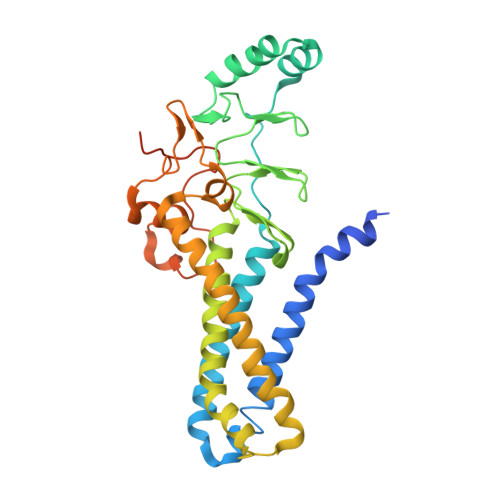
7KHM

Crystal structure of hDHHS20 bound to palmitoyl CoA


Experimental Data Snapshot

  • Method: X-RAY DIFFRACTION
  • Resolution: 2.88 Å
  • R-Value Free: 0.325 
  • R-Value Work: 0.294 
  • R-Value Observed: 0.296 

Starting Model: experimental
View more details

wwPDB Validation   3D Report Full Report


Ligand Structure Quality Assessment 


This is version 1.2 of the entry. See complete history


Literature

Bivalent recognition of fatty acyl-CoA by a human integral membrane palmitoyltransferase.

Lee, C.J.Stix, R.Rana, M.S.Shikwana, F.Murphy, R.E.Ghirlando, R.Faraldo-Gomez, J.D.Banerjee, A.

(2022) Proc Natl Acad Sci U S A 119

  • DOI: https://doi.org/10.1073/pnas.2022050119
  • Primary Citation of Related Structures:  
    7KHM

  • PubMed Abstract: 

    S-acylation, also known as palmitoylation, is the most abundant form of protein lipidation in humans. This reversible posttranslational modification, which targets thousands of proteins, is catalyzed by 23 members of the DHHC family of integral membrane enzymes. DHHC enzymes use fatty acyl-CoA as the ubiquitous fatty acyl donor and become autoacylated at a catalytic cysteine; this intermediate subsequently transfers the fatty acyl group to a cysteine in the target protein. Protein S-acylation intersects with almost all areas of human physiology, and several DHHC enzymes are considered as possible therapeutic targets against diseases such as cancer. These efforts would greatly benefit from a detailed understanding of the molecular basis for this crucial enzymatic reaction. Here, we combine X-ray crystallography with all-atom molecular dynamics simulations to elucidate the structure of the precatalytic complex of human DHHC20 in complex with palmitoyl CoA. The resulting structure reveals that the fatty acyl chain inserts into a hydrophobic pocket within the transmembrane spanning region of the protein, whereas the CoA headgroup is recognized by the cytosolic domain through polar and ionic interactions. Biochemical experiments corroborate the predictions from our structural model. We show, using both computational and experimental analyses, that palmitoyl CoA acts as a bivalent ligand where the interaction of the DHHC enzyme with both the fatty acyl chain and the CoA headgroup is important for catalytic chemistry to proceed. This bivalency explains how, in the presence of high concentrations of free CoA under physiological conditions, DHHC enzymes can efficiently use palmitoyl CoA as a substrate for autoacylation.


  • Organizational Affiliation

    Section on Structural and Chemical Biology of Membrane Proteins, Eunice Kennedy Shriver National Institute of Child Health and Human Development, NIH, Bethesda, MD 20892.


Macromolecules
Find similar proteins by:  (by identity cutoff)  |  3D Structure
Entity ID: 1
MoleculeChains Sequence LengthOrganismDetailsImage
Isoform 4 of Palmitoyltransferase ZDHHC20
A, B
330Homo sapiensMutation(s): 1 
Gene Names: ZDHHC20
EC: 2.3.1.225 (PDB Primary Data), 2.3.1 (PDB Primary Data)
Membrane Entity: Yes 
UniProt & NIH Common Fund Data Resources
Find proteins for Q5W0Z9 (Homo sapiens)
Explore Q5W0Z9 
Go to UniProtKB:  Q5W0Z9
PHAROS:  Q5W0Z9
GTEx:  ENSG00000180776 
Entity Groups  
Sequence Clusters30% Identity50% Identity70% Identity90% Identity95% Identity100% Identity
UniProt GroupQ5W0Z9
Sequence Annotations
Expand
  • Reference Sequence
Small Molecules
Ligands 3 Unique
IDChains Name / Formula / InChI Key2D Diagram3D Interactions
PKZ (Subject of Investigation/LOI)
Query on PKZ

Download Ideal Coordinates CCD File 
E [auth A],
I [auth B]
Palmitoyl-CoA
C37 H66 N7 O17 P3 S
MNBKLUUYKPBKDU-BBECNAHFSA-N
PO4
Query on PO4

Download Ideal Coordinates CCD File 
F [auth A]PHOSPHATE ION
O4 P
NBIIXXVUZAFLBC-UHFFFAOYSA-K
ZN
Query on ZN

Download Ideal Coordinates CCD File 
C [auth A],
D [auth A],
G [auth B],
H [auth B]
ZINC ION
Zn
PTFCDOFLOPIGGS-UHFFFAOYSA-N
Experimental Data & Validation

Experimental Data

  • Method: X-RAY DIFFRACTION
  • Resolution: 2.88 Å
  • R-Value Free: 0.325 
  • R-Value Work: 0.294 
  • R-Value Observed: 0.296 
  • Space Group: P 1 21 1
Unit Cell:
Length ( Å )Angle ( ˚ )
a = 53.529α = 90
b = 114.61β = 91.23
c = 83.596γ = 90
Software Package:
Software NamePurpose
PHENIXrefinement
XDSdata reduction
Aimlessdata scaling
PHASERphasing
PDB_EXTRACTdata extraction

Structure Validation

View Full Validation Report


Ligand Structure Quality Assessment 


Entry History 

Deposition Data

Revision History  (Full details and data files)

  • Version 1.0: 2022-02-02
    Type: Initial release
  • Version 1.1: 2022-02-23
    Changes: Database references
  • Version 1.2: 2023-10-18
    Changes: Data collection, Refinement description