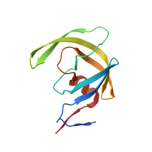
Revertant mutation V48G alters conformational dynamics of highly drug resistant HIV protease PRS17.
Burnaman, S.H., Kneller, D.W., Wang, Y.F., Kovalevsky, A., Weber, I.T.(2021) J Mol Graph Model 108: 108005-108005
- PubMed: 34419931
- DOI: https://doi.org/10.1016/j.jmgm.2021.108005
- Primary Citation of Related Structures:
7N6T, 7N6V, 7N6X - PubMed Abstract:
Drug resistance is a serious problem for controlling the HIV/AIDS pandemic. Current antiviral drugs show several orders of magnitude worse inhibition of highly resistant clinical variant PRS17 of HIV-1 protease compared with wild-type protease. We have analyzed the effects of a common resistance mutation G48V in the flexible flaps of the protease by assessing the revertant PRS17 V48G for changes in enzyme kinetics, inhibition, structure, and dynamics. Both PRS17 and the revertant showed about 10-fold poorer catalytic efficiency than wild-type enzyme (0.55 and 0.39 μM -1 min -1 compared to 6.3 μM -1 min -1 ). Clinical inhibitors, amprenavir and darunavir, showed 2-fold and 8-fold better inhibition, respectively, of the revertant than of PRS17, although the inhibition constants for PRS17 V48G were still 25 to 1,200-fold worse than for wild-type protease. Crystal structures of inhibitor-free revertant and amprenavir complexes with revertant and PRS17 were solved at 1.3-1.5 Å resolution. The amprenavir complexes of PRS17 V48G and PRS17 showed no significant differences in the interactions with inhibitor, although changes were observed in the conformation of Phe53 and the interactions of the flaps. The inhibitor-free structure of the revertant showed flaps in an open conformation, however, the flap tips do not have the unusual curled conformation seen in inhibitor-free PRS17. Molecular dynamics simulations were run for 1 μs on the two inhibitor-free mutants and wild-type protease. PRS17 exhibited higher conformational fluctuations than the revertant, while the wild-type protease adopted the closed conformation and showed the least variation. The second half of the simulations captured the transition of the flaps of PRS17 from a closed to a semi-open state, whereas the flaps of PRS17 V48G tucked into the active site and the wild-type protease retained the closed conformation. These results suggest that mutation G48V contributes to drug resistance by altering the conformational dynamics of the flaps.
- Department of Biology, Georgia State University, Atlanta, GA, USA.
Organizational Affiliation:


