7ZUH | pdb_00007zuh

PENICILLIN-BINDING PROTEIN 1B (PBP-1B) Streptococcus pneumoniae R6


Experimental Data Snapshot

  • Method: X-RAY DIFFRACTION
  • Resolution: 1.47 Å
  • R-Value Free: 
    0.200 (Depositor), 0.200 (DCC) 
  • R-Value Work: 
    0.189 (Depositor), 0.180 (DCC) 

Starting Model: experimental
View more details

wwPDB Validation   3D Report Full Report


This is version 1.3 of the entry. See complete history

Re-refinement Note

A newer entry is available that reflects an alternative modeling of the original data: 7ZUI 7ZUJ 7ZUK 7ZUL


Literature

Structural and Dynamics Analyses of beta-Lactam Inhibition of Streptococcus pneumoniae Penicillin-Binding Protein 1b (PBP1b) Guide Interrogation of Structure-Activity Relationships.

Flanders, P.L.Gillingham, J.R.Contreras-Martel, C.Dessen, A.Carlson, E.E.Ambrose, E.A.

(2026) ACS Chem Biol 

  • DOI: https://doi.org/10.1021/acschembio.5c00788
  • Primary Citation of Related Structures:  
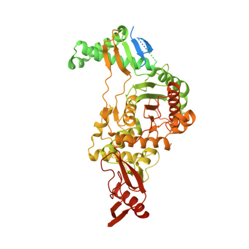
    7ZUH, 9SSD, 9SSE, 9SSF, 9SSG, 9SSH, 9SSI

  • PubMed Abstract: 

    The Gram-positive pathogen Streptococcus pneumoniae , like the majority of bacteria, contains a peptidoglycan-based cell wall whose structure is highly dependent on the action of penicillin-binding proteins (PBPs). While the β-lactam antibiotics have been employed as an antimicrobial strategy for nearly a century, much remains unclear about how inhibitor structure informs potency and PBP isoform selectivity. Here, we obtained high-resolution structures (<2Å) of S. pneumoniae PBP1b cocrystallized with 6 β-lactams. Surprisingly, 2 structures feature a noncanonical conformation of the covalent "acyl-enzyme complex." To clarify how protein-ligand interactions mediate inhibitor binding, we applied molecular modeling and molecular mechanics-based dynamics analyses. Our analyses illustrate how seemingly minimal changes to inhibitor structure modulate β-lactam binding mode and inhibitor potency, as described by the metric k inact / K I . Furthermore, we demonstrate that persistent interaction in the covalent acyl-enzyme complex between the inhibitor carboxylate and a highly conserved three-residue motif is not fully predictive of k inact / K I for PBP1b. In silico modeling suggests that the noncovalent preacyl complex may leverage this interaction, but a postacylation change in ligand conformation may accompany acylation in some inhibitors. The elucidation of key PBP1b ligand-receptor interactions pre- and postacylation will inform the rational design of novel PBP inhibitors and probes.


  • Organizational Affiliation
    • Department of Medicinal Chemistry, University of Minnesota, 208 Harvard Street SE, Minneapolis, Minnesota 55454, United States.

Macromolecules
Find similar proteins by:  (by identity cutoff)  |  3D Structure
Entity ID: 1
MoleculeChains Sequence LengthOrganismDetailsImage
Penicillin-binding protein 1bA [auth AAA]821Streptococcus pneumoniae R6Mutation(s): 3 
Gene Names: pbp1bspr1909
EC: 2.3.2 (PDB Primary Data), 2.4.1.129 (PDB Primary Data)
UniProt
Find proteins for Q7CRA4 (Streptococcus pneumoniae (strain ATCC BAA-255 / R6))
Explore Q7CRA4 
Go to UniProtKB:  Q7CRA4
Entity Groups  
Sequence Clusters30% Identity50% Identity70% Identity90% Identity95% Identity100% Identity
UniProt GroupQ7CRA4
Sequence Annotations
Expand
  • Reference Sequence
Small Molecules
Ligands 2 Unique
IDChains Name / Formula / InChI Key2D Diagram3D Interactions
CL
Query on CL

Download Ideal Coordinates CCD File 
AA [auth AAA]
C [auth AAA]
D [auth AAA]
E [auth AAA]
F [auth AAA]
AA [auth AAA],
C [auth AAA],
D [auth AAA],
E [auth AAA],
F [auth AAA],
G [auth AAA],
H [auth AAA],
I [auth AAA],
J [auth AAA],
K [auth AAA],
L [auth AAA],
M [auth AAA],
N [auth AAA],
O [auth AAA],
P [auth AAA],
Q [auth AAA],
R [auth AAA],
S [auth AAA],
T [auth AAA],
U [auth AAA],
V [auth AAA],
W [auth AAA],
X [auth AAA],
Y [auth AAA],
Z [auth AAA]
CHLORIDE ION
Cl
VEXZGXHMUGYJMC-UHFFFAOYSA-M
MG
Query on MG

Download Ideal Coordinates CCD File 
B [auth AAA]MAGNESIUM ION
Mg
JLVVSXFLKOJNIY-UHFFFAOYSA-N
Experimental Data & Validation

Experimental Data

  • Method: X-RAY DIFFRACTION
  • Resolution: 1.47 Å
  • R-Value Free:  0.200 (Depositor), 0.200 (DCC) 
  • R-Value Work:  0.189 (Depositor), 0.180 (DCC) 
Space Group: C 2 2 21
Unit Cell:
Length ( Å )Angle ( ˚ )
a = 95.681α = 90
b = 147.016β = 90
c = 98.709γ = 90
Software Package:
Software NamePurpose
REFMACrefinement
XDSdata reduction
XSCALEdata scaling
PHASERphasing

Structure Validation

View Full Validation Report


Entry History & Funding Information

Deposition Data


Funding OrganizationLocationGrant Number
Agence Nationale de la Recherche (ANR)FranceANR-10-INBS-0005-02
Agence Nationale de la Recherche (ANR)FranceANR-17-EURE-0003
National Institutes of Health/National Center for Research Resources (NIH/NCRR)United StatesR01 GM128439
National Institutes of Health/National Center for Research Resources (NIH/NCRR)United StatesK12 GM119955
National Institutes of Health/National Center for Research Resources (NIH/NCRR)United StatesTL1R002493
National Institutes of Health/National Center for Research Resources (NIH/NCRR)United StatesUL1TR002494

Revision History  (Full details and data files)

  • Version 1.0: 2022-11-02
    Type: Initial release
  • Version 1.1: 2022-11-30
    Changes: Database references
  • Version 1.2: 2024-01-31
    Changes: Data collection, Refinement description
  • Version 1.3: 2026-02-18
    Changes: Database references, Structure summary