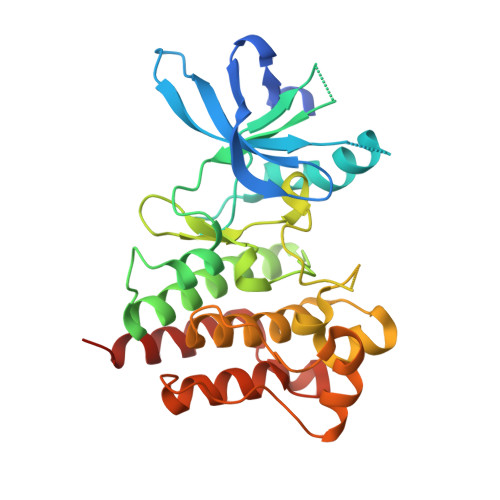
Biophysical and structural characterization of the impacts of MET phosphorylation on tepotinib binding.
Gradler, U., Schwarz, D., Wegener, A., Eichhorn, T., Bandeiras, T.M., Freitas, M.C., Lammens, A., Ganichkin, O., Augustin, M., Minguzzi, S., Becker, F., Bomke, J.(2023) J Biological Chem 299: 105328-105328
- PubMed: 37806493
- DOI: https://doi.org/10.1016/j.jbc.2023.105328
- Primary Citation of Related Structures:
8AU3, 8AU5, 8AW1 - PubMed Abstract:
The receptor tyrosine kinase MET is activated by hepatocyte growth factor binding, followed by phosphorylation of the intracellular kinase domain (KD) mainly within the activation loop (A-loop) on Y1234 and Y1235. Dysregulation of MET can lead to both tumor growth and metastatic progression of cancer cells. Tepotinib is a highly selective, potent type Ib MET inhibitor and approved for treatment of non-small cell lung cancer harboring METex14 skipping alterations. Tepotinib binds to the ATP site of unphosphorylated MET with critical π-stacking contacts to Y1230 of the A-loop, resulting in a high residence time. In our study, we combined protein crystallography, biophysical methods (surface plasmon resonance, differential scanning fluorimetry), and mass spectrometry to clarify the impacts of A-loop conformation on tepotinib binding using different recombinant MET KD protein variants. We solved the first crystal structures of MET mutants Y1235D, Y1234E/1235E, and F1200I in complex with tepotinib. Our biophysical and structural data indicated a linkage between reduced residence times for tepotinib and modulation of A-loop conformation either by mutation (Y1235D), by affecting the overall Y1234/Y1235 phosphorylation status (L1195V and F1200I) or by disturbing critical π-stacking interactions with tepotinib (Y1230C). We corroborated these data with target engagement studies by fluorescence cross-correlation spectroscopy using KD constructs in cell lysates or full-length receptors from solubilized cellular membranes as WT or activated mutants (Y1235D and Y1234E/1235E). Collectively, our results provide further insight into the MET A-loop structural determinants that affect the binding of the selective inhibitor tepotinib.
- The Healthcare Business of Merck KGaA, Darmstadt, Germany. Electronic address: ulrich.graedler@emdserono.com.
Organizational Affiliation:


