


Structure of AcuB from Bacillus subtilis
Zheng, L.J., Bange, G.To be published.
Experimental Data Snapshot
Starting Model: in silico
View more details
Entity ID: 1 | |||||
|---|---|---|---|---|---|
| Molecule | Chains | Sequence Length | Organism | Details | Image |
| Acetoin utilization protein AcuB | 215 | Bacillus subtilis subsp. subtilis str. 168 | Mutation(s): 0 Gene Names: acuB, BSU29700 | 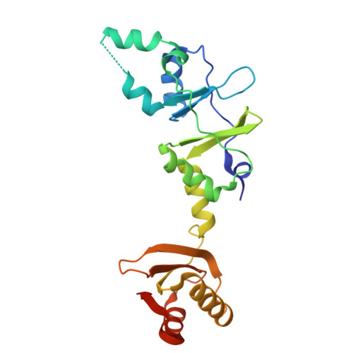 | |
UniProt | |||||
Find proteins for P39066 (Bacillus subtilis (strain 168)) Explore P39066 Go to UniProtKB: P39066 | |||||
Entity Groups | |||||
| Sequence Clusters | 30% Identity50% Identity70% Identity90% Identity95% Identity100% Identity | ||||
| UniProt Group | P39066 | ||||
Sequence AnnotationsExpand | |||||
| |||||
| Ligands 1 Unique | |||||
|---|---|---|---|---|---|
| ID | Chains | Name / Formula / InChI Key | 2D Diagram | 3D Interactions | |
| B4P (Subject of Investigation/LOI) Query on B4P | C [auth A], D [auth B] | BIS(ADENOSINE)-5'-TETRAPHOSPHATE C20 H28 N10 O19 P4 YOAHKNVSNCMZGQ-XPWFQUROSA-N |  | ||
| Length ( Å ) | Angle ( ˚ ) |
|---|---|
| a = 81.546 | α = 90 |
| b = 81.546 | β = 90 |
| c = 338.549 | γ = 120 |
| Software Name | Purpose |
|---|---|
| PHENIX | refinement |
| PHENIX | phasing |
| autoPROC | data processing |
| XDS | data scaling |
| autoPROC | data reduction |
| Funding Organization | Location | Grant Number |
|---|---|---|
| Max Planck Society | Germany | -- |


