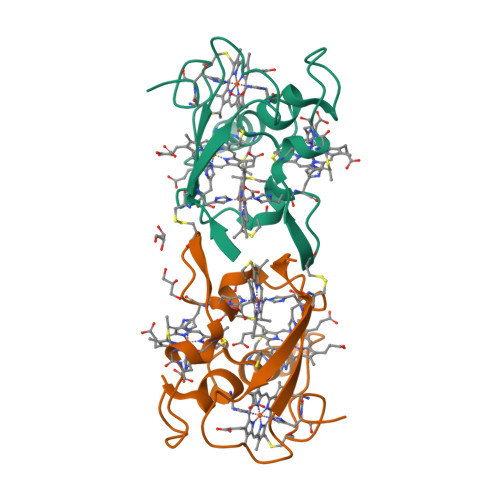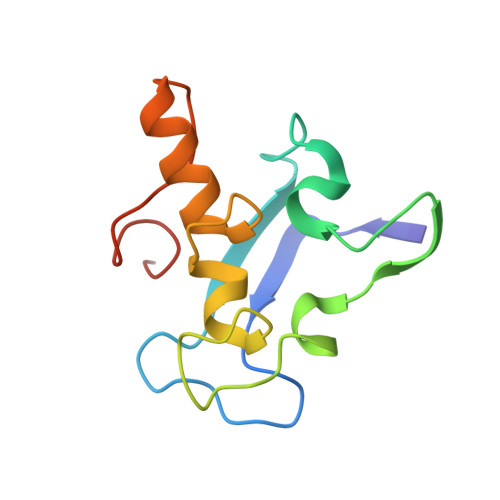
Structure of Dimeric Cytochrome C3 from Desulfovibrio Gigas at 1.2 A Resolution
Aragao, D., Frazao, C., Sieker, L., Sheldrick, G.M., Legall, J., Carrondo, M.A.(2003) Acta Crystallogr D Biol Crystallogr 59: 644
- PubMed: 12657783
- DOI: https://doi.org/10.1107/s090744490300194x
- Primary Citation of Related Structures:
1GYO - PubMed Abstract:
The structure of dimeric cytochrome c(3) from the sulfate-reducing bacterium Desulfovibrio gigas, diDg, obtained by ab initio methods was further refined to 1.2 A resolution, giving final reliability factors of R(free) = 14.8% and R = 12.4%. This cytochrome is a dimer of tetraheme cytochrome c(3) molecules covalently linked by two solvent-accessible disulfide bridges, a characteristic unique to members of the cytochrome c(3) superfamily. Anisotropic analysis using the semi-rigid TLS method shows different behaviour for analogous loops in each monomer arising from their different packing environments. A detailed sequence and structural comparison with all other known cytochrome c(3) domains in single- and multi-domain cytochromes c(3) shows the presence of structurally conserved regions in this family, despite the high variability of the amino-acid sequence. An internal water molecule is conserved in a common structural arrangement in all c(3) tetraheme domains, indicating a probable electron-transfer pathway between hemes I and II. Unique features of diDg are an internal methionine residue close to heme I and to one of the axial ligands of heme III, where all other structures of the cytochrome c(3) superfamily have a phenylalanine, and a rather unusual CXXXCH heme-binding motif only found so far in this cytochrome.
Organizational Affiliation:
Instituto de Tecnologia Química e Biológica, Oeiras, Portugal.