1OIX | pdb_00001oix

X-ray structure of the small G protein Rab11a in complex with GDP and Pi


Experimental Data Snapshot

  • Method: X-RAY DIFFRACTION
  • Resolution: 1.70 Å
  • R-Value Free: 
    0.229 (Depositor), 0.220 (DCC) 
  • R-Value Work: 
    0.201 (Depositor), 0.200 (DCC) 
  • R-Value Observed: 
    0.201 (Depositor) 

Starting Model: experimental
View more details

wwPDB Validation   3D Report Full Report


Ligand Structure Quality Assessment 

Created with Raphaël 2.3.0Worse 01 BetterLigand structure goodness of fit to experimental dataBest fitted GDPClick on this verticalbar to view details

This is version 1.3 of the entry. See complete history


Literature

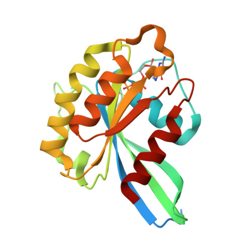
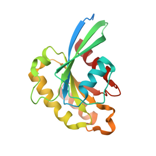
Crystallographic Evidence for Substrate-Assisted GTP Hydrolysis by a Small GTP Binding Protein

Pasqualato, S.Cherfils, J.

(2005) Structure 13: 533

  • DOI: https://doi.org/10.1016/j.str.2005.01.014
  • Primary Citation of Related Structures:  
    1OIX

  • PubMed Abstract: 

    GTP hydrolysis by small GTP binding proteins of the Ras superfamily is a universal reaction that controls multiple cellular regulations. Its enzymic mechanism has been the subject of long-standing debates as to the existence/identity of the general base and the electronic nature of its transition state. Here we report the high-resolution crystal structure of a small GTP binding protein, Rab11, solved in complex with GDP and Pi. Unexpectedly, a Pi oxygen and the GDP-cleaved oxygen are located less than 2.5 A apart, suggesting that they share a proton, likely in the form of a low-barrier hydrogen bond. This implies that the gamma-phosphate of GTP was protonated; hence, that GTP acts as a general base. Furthermore, this interaction should establish at, and stabilize, the transition state. Altogether, we propose a revised model for the GTPase reaction that should reconcile earlier models into a unique substrate-assisted mechanism.


  • Organizational Affiliation

    Laboratoire d'Enzymologie et Biochimie Structurales, CNRS, Avenue de la Terrasse, 91198 Gif-sur-Yvette Cedex, France.


Macromolecules
Find similar proteins by:  (by identity cutoff)  |  3D Structure
Entity ID: 1
MoleculeChains Sequence LengthOrganismDetailsImage
RAS-RELATED PROTEIN RAB-11A191Homo sapiensMutation(s): 1 
EC: 3.6.5.2
UniProt & NIH Common Fund Data Resources
Find proteins for P62491 (Homo sapiens)
Explore P62491 
Go to UniProtKB:  P62491
PHAROS:  P62491
GTEx:  ENSG00000103769 
Entity Groups  
Sequence Clusters30% Identity50% Identity70% Identity90% Identity95% Identity100% Identity
UniProt GroupP62491
Sequence Annotations
Expand
  • Reference Sequence
Experimental Data & Validation

Experimental Data

  • Method: X-RAY DIFFRACTION
  • Resolution: 1.70 Å
  • R-Value Free:  0.229 (Depositor), 0.220 (DCC) 
  • R-Value Work:  0.201 (Depositor), 0.200 (DCC) 
  • R-Value Observed: 0.201 (Depositor) 
Space Group: I 4 2 2
Unit Cell:
Length ( Å )Angle ( ˚ )
a = 74.077α = 90
b = 74.077β = 90
c = 124.905γ = 90
Software Package:
Software NamePurpose
CNSrefinement
DENZOdata reduction
SCALEPACKdata scaling
AMoREphasing

Structure Validation

View Full Validation Report


Ligand Structure Quality Assessment 

Created with Raphaël 2.3.0Worse 01 BetterLigand structure goodness of fit to experimental dataBest fitted GDPClick on this verticalbar to view details

Entry History 

Deposition Data

Revision History  (Full details and data files)

  • Version 1.0: 2005-01-25
    Type: Initial release
  • Version 1.1: 2011-05-08
    Changes: Version format compliance
  • Version 1.2: 2011-07-13
    Changes: Version format compliance
  • Version 1.3: 2023-12-13
    Changes: Data collection, Database references, Derived calculations, Other, Refinement description