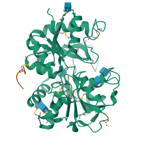
Crystal Structure of C-lobe of Bovine lactoferrin Complexed with N-acetylmuramyl l-alanyl d-isoglutamine at 1.8 A Resolution
Shukla, P.K., Gautam, L., Sinha, M., Kaur, P., Sharma, S., Singh, T.P.To be published.
Experimental Data Snapshot
Starting Model: experimental
View more details
Entity ID: 1 | |||||
|---|---|---|---|---|---|
| Molecule | Chains | Sequence Length | Organism | Details | Image |
| Lactotransferrin | 335 | Bos taurus | Mutation(s): 0 EC: 3.4.21 | 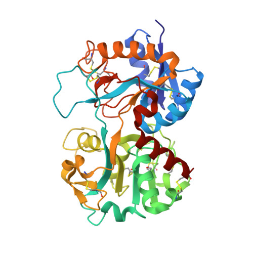 | |
UniProt | |||||
Find proteins for P24627 (Bos taurus) Explore P24627 Go to UniProtKB: P24627 | |||||
Entity Groups | |||||
| Sequence Clusters | 30% Identity50% Identity70% Identity90% Identity95% Identity100% Identity | ||||
| UniProt Group | P24627 | ||||
Glycosylation | |||||
| Glycosylation Sites: 3 | |||||
Sequence AnnotationsExpand | |||||
| |||||
Find similar proteins by: Sequence | 3D Structure
| Ligands 9 Unique | |||||
|---|---|---|---|---|---|
| ID | Chains | Name / Formula / InChI Key | 2D Diagram | 3D Interactions | |
| NAG Query on NAG | E [auth A] | 2-acetamido-2-deoxy-beta-D-glucopyranose C8 H15 N O6 OVRNDRQMDRJTHS-FMDGEEDCSA-N |  | ||
| NDG Query on NDG | N [auth A] | 2-acetamido-2-deoxy-alpha-D-glucopyranose C8 H15 N O6 OVRNDRQMDRJTHS-PVFLNQBWSA-N |  | ||
| DGN Query on DGN | K [auth A] | D-GLUTAMINE C5 H10 N2 O3 ZDXPYRJPNDTMRX-GSVOUGTGSA-N |  | ||
| SO4 Query on SO4 | J [auth A] | SULFATE ION O4 S QAOWNCQODCNURD-UHFFFAOYSA-L |  | ||
| LAC Query on LAC | M [auth A] | LACTIC ACID C3 H6 O3 JVTAAEKCZFNVCJ-UWTATZPHSA-N |  | ||
| ALA Query on ALA | L [auth A] | ALANINE C3 H7 N O2 QNAYBMKLOCPYGJ-REOHCLBHSA-N |  | ||
| ZN Query on ZN | G [auth A], H [auth A] | ZINC ION Zn PTFCDOFLOPIGGS-UHFFFAOYSA-N |  | ||
| CO3 Query on CO3 | I [auth A] | CARBONATE ION C O3 BVKZGUZCCUSVTD-UHFFFAOYSA-L |  | ||
| FE Query on FE | F [auth A] | FE (III) ION Fe VTLYFUHAOXGGBS-UHFFFAOYSA-N |  | ||
| Length ( Å ) | Angle ( ˚ ) |
|---|---|
| a = 62.55 | α = 90 |
| b = 49.951 | β = 107.03 |
| c = 65.129 | γ = 90 |
| Software Name | Purpose |
|---|---|
| HKL-2000 | data collection |
| AMoRE | phasing |
| REFMAC | refinement |
| DENZO | data reduction |
| SCALEPACK | data scaling |
RCSB PDB is hosted by

RCSB PDB is a member of the