1RGI | pdb_00001rgi

Crystal structure of gelsolin domains G1-G3 bound to actin


Experimental Data Snapshot

  • Method: X-RAY DIFFRACTION
  • Resolution: 3.00 Å
  • R-Value Free: 
    0.258 (Depositor), 0.220 (DCC) 
  • R-Value Work: 
    0.224 (Depositor), 0.190 (DCC) 
  • R-Value Observed: 
    0.226 (Depositor) 

Starting Model: experimental
View more details

wwPDB Validation   3D Report Full Report


Ligand Structure Quality Assessment 

Created with Raphaël 2.3.0Worse 01 BetterLigand structure goodness of fit to experimental dataBest fitted ATPClick on this verticalbar to view details

This is version 1.4 of the entry. See complete history


Literature

Structure of the N-terminal half of gelsolin bound to actin: roles in severing, apoptosis and FAF

Burtnick, L.D.Urosev, D.Irobi, E.Narayan, K.Robinson, R.C.

(2004) EMBO J 23: 2713-2722

  • DOI: https://doi.org/10.1038/sj.emboj.7600280
  • Primary Citation of Related Structures:  
    1RGI

  • PubMed Abstract: 

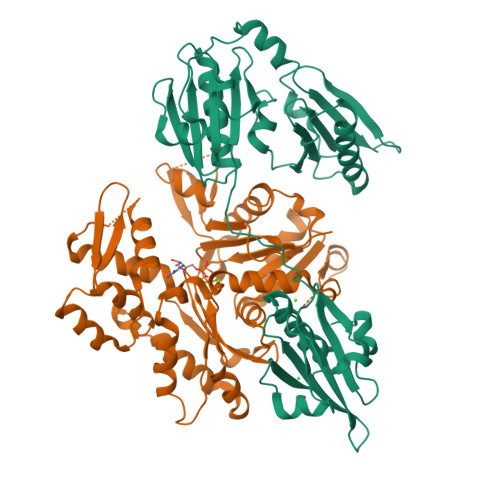
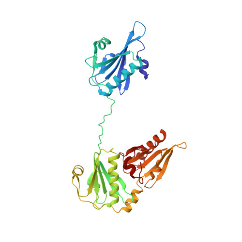
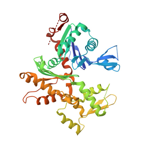
    The actin filament-severing functionality of gelsolin resides in its N-terminal three domains (G1-G3). We have determined the structure of this fragment in complex with an actin monomer. The structure reveals the dramatic domain rearrangements that activate G1-G3, which include the replacement of interdomain interactions observed in the inactive, calcium-free protein by new contacts to actin, and by a novel G2-G3 interface. Together, these conformational changes are critical for actin filament severing, and we suggest that their absence leads to the disease Finnish-type familial amyloidosis. Furthermore, we propose that association with actin drives the calcium-independent activation of isolated G1-G3 during apoptosis, and that a similar mechanism operates to activate native gelsolin at micromolar levels of calcium. This is the first structure of a filament-binding protein bound to actin and it sets stringent, high-resolution limitations on the arrangement of actin protomers within the filament.


  • Organizational Affiliation

    Department of Chemistry and Centre for Blood Research, The University of British Columbia, Vancouver, BC, Canada.


Macromolecules
Find similar proteins by:  (by identity cutoff)  |  3D Structure
Entity ID: 1
MoleculeChains Sequence LengthOrganismDetailsImage
GelsolinA [auth G]346Equus caballusMutation(s): 0 
UniProt
Find proteins for Q28372 (Equus caballus)
Explore Q28372 
Go to UniProtKB:  Q28372
Entity Groups  
Sequence Clusters30% Identity50% Identity70% Identity90% Identity95% Identity100% Identity
UniProt GroupQ28372
Sequence Annotations
Expand
  • Reference Sequence
Find similar proteins by:  (by identity cutoff)  |  3D Structure
Entity ID: 2
MoleculeChains Sequence LengthOrganismDetailsImage
Actin, alpha skeletal muscleB [auth A]377Oryctolagus cuniculusMutation(s): 0 
EC: 3.6.4
UniProt
Find proteins for P68135 (Oryctolagus cuniculus)
Explore P68135 
Go to UniProtKB:  P68135
Entity Groups  
Sequence Clusters30% Identity50% Identity70% Identity90% Identity95% Identity100% Identity
UniProt GroupP68135
Sequence Annotations
Expand
  • Reference Sequence
Experimental Data & Validation

Experimental Data

  • Method: X-RAY DIFFRACTION
  • Resolution: 3.00 Å
  • R-Value Free:  0.258 (Depositor), 0.220 (DCC) 
  • R-Value Work:  0.224 (Depositor), 0.190 (DCC) 
  • R-Value Observed: 0.226 (Depositor) 
Space Group: P 31 2 1
Unit Cell:
Length ( Å )Angle ( ˚ )
a = 145.245α = 90
b = 145.245β = 90
c = 129.948γ = 120
Software Package:
Software NamePurpose
REFMACrefinement
SCALEPACKdata scaling
AMoREphasing
CNSrefinement
DENZOdata reduction

Structure Validation

View Full Validation Report


Ligand Structure Quality Assessment 

Created with Raphaël 2.3.0Worse 01 BetterLigand structure goodness of fit to experimental dataBest fitted ATPClick on this verticalbar to view details

Entry History 

Deposition Data

Revision History  (Full details and data files)

  • Version 1.0: 2004-07-27
    Type: Initial release
  • Version 1.1: 2008-04-29
    Changes: Version format compliance
  • Version 1.2: 2011-07-13
    Changes: Advisory, Version format compliance
  • Version 1.3: 2019-07-24
    Changes: Data collection, Refinement description
  • Version 1.4: 2023-08-23
    Changes: Data collection, Database references, Derived calculations, Refinement description