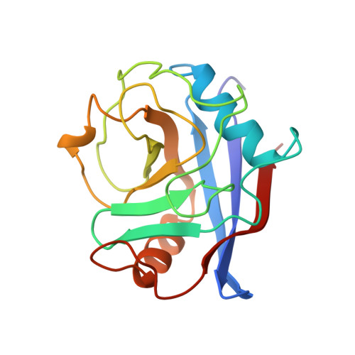
1AK4

HUMAN CYCLOPHILIN A BOUND TO THE AMINO-TERMINAL DOMAIN OF HIV-1 CAPSID


Experimental Data Snapshot

  • Method: X-RAY DIFFRACTION
  • Resolution: 2.36 Å
  • R-Value Free: 0.306 
  • R-Value Work: 0.238 
  • R-Value Observed: 0.238 

Starting Model: experimental
View more details

wwPDB Validation   3D Report Full Report


This is version 1.4 of the entry. See complete history


Literature

Crystal structure of human cyclophilin A bound to the amino-terminal domain of HIV-1 capsid.

Gamble, T.R.Vajdos, F.F.Yoo, S.Worthylake, D.K.Houseweart, M.Sundquist, W.I.Hill, C.P.

(1996) Cell 87: 1285-1294

  • DOI: https://doi.org/10.1016/s0092-8674(00)81823-1
  • Primary Citation of Related Structures:  
    1AK4

  • PubMed Abstract: 

    The HIV-1 capsid protein forms the conical core structure at the center of the mature virion. Capsid also binds the human peptidyl prolyl isomerase, cyclophilin A, thereby packaging the enzyme into the virion. Cyclophilin A subsequently performs an essential function in HIV-1 replication, possibly helping to disassemble the capsid core upon infection. We report the 2.36 A crystal structure of the N-terminal domain of HIV-1 capsid (residues 1-151) in complex with human cyclophilin A. A single exposed capsid loop (residues 85-93) binds in the enzyme's active site, and Pro-90 adopts an unprecedented trans conformation. The structure suggests how cyclophilin A can act as a sequence-specific binding protein and a nonspecific prolyl isomerase. In the crystal lattice, capsid molecules assemble into continuous planar strips. Side by side association of these strips may allow capsid to form the surface of the viral core. Cyclophilin A could then function by weakening the association between capsid strips, thereby promoting disassembly of the viral core.


  • Organizational Affiliation

    Biochemistry Department, University of Utah, Salt Lake City 84103, USA.


Macromolecules
Find similar proteins by:  (by identity cutoff)  |  3D Structure
Entity ID: 1
MoleculeChains Sequence LengthOrganismDetailsImage
CYCLOPHILIN A
A, B
165Homo sapiensMutation(s): 0 
Gene Names: CYCLOPHILIN
EC: 5.2.1.8
UniProt & NIH Common Fund Data Resources
Find proteins for P62937 (Homo sapiens)
Explore P62937 
Go to UniProtKB:  P62937
PHAROS:  P62937
GTEx:  ENSG00000196262 
Entity Groups  
Sequence Clusters30% Identity50% Identity70% Identity90% Identity95% Identity100% Identity
UniProt GroupP62937
Sequence Annotations
Expand
  • Reference Sequence
Find similar proteins by:  (by identity cutoff)  |  3D Structure
Entity ID: 2
MoleculeChains Sequence LengthOrganismDetailsImage
HIV-1 CAPSID
C, D
145Human immunodeficiency virus 1Mutation(s): 0 
Gene Names: CYCLOPHILIN
UniProt
Find proteins for P12497 (Human immunodeficiency virus type 1 group M subtype B (isolate NY5))
Explore P12497 
Go to UniProtKB:  P12497
Entity Groups  
Sequence Clusters30% Identity50% Identity70% Identity90% Identity95% Identity100% Identity
UniProt GroupP12497
Sequence Annotations
Expand
  • Reference Sequence
Experimental Data & Validation

Experimental Data

  • Method: X-RAY DIFFRACTION
  • Resolution: 2.36 Å
  • R-Value Free: 0.306 
  • R-Value Work: 0.238 
  • R-Value Observed: 0.238 
  • Space Group: P 1 21 1
Unit Cell:
Length ( Å )Angle ( ˚ )
a = 38.6α = 90
b = 113.1β = 100.7
c = 67γ = 90
Software Package:
Software NamePurpose
X-PLORmodel building
X-PLORrefinement
DENZOdata reduction
SCALEPACKdata scaling
X-PLORphasing

Structure Validation

View Full Validation Report


Entry History 

Deposition Data

Revision History  (Full details and data files)

  • Version 1.0: 1997-10-15
    Type: Initial release
  • Version 1.1: 2008-03-24
    Changes: Version format compliance
  • Version 1.2: 2011-07-13
    Changes: Version format compliance
  • Version 1.3: 2023-08-02
    Changes: Database references, Other, Refinement description
  • Version 1.4: 2024-05-22
    Changes: Data collection