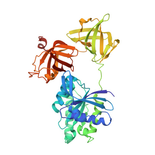
1D2E | pdb_00001d2e

CRYSTAL STRUCTURE OF MITOCHONDRIAL EF-TU IN COMPLEX WITH GDP


Experimental Data Snapshot

  • Method: X-RAY DIFFRACTION
  • Resolution: 1.94 Å
  • R-Value Free: 
    0.257 (Depositor) 
  • R-Value Work: 
    0.237 (Depositor), 0.240 (DCC) 
  • R-Value Observed: 
    0.237 (Depositor) 

wwPDB Validation   3D Report Full Report


Ligand Structure Quality Assessment 

Created with Raphaël 2.3.0Worse 01 BetterLigand structure goodness of fit to experimental dataBest fitted GDPClick on this verticalbar to view details

This is version 1.4 of the entry. See complete history


Literature

High resolution crystal structure of bovine mitochondrial EF-Tu in complex with GDP.

Andersen, G.R.Thirup, S.Spremulli, L.L.Nyborg, J.

(2000) J Mol Biology 297: 421-436

  • DOI: https://doi.org/10.1006/jmbi.2000.3564
  • Primary Citation of Related Structures:  
    1D2E

  • PubMed Abstract: 

    The crystal structure of bovine mitochondrial elongation factor Tu (EF-Tu) in complex with GDP has been determined at a resolution of 1. 94 A. The structure is similar to that of EF-Tu:GDP from Escherichia coli and Thermus aquaticus, but the orientation of the GDP-binding domain 1 is changed relative to domains 2 and 3. Sixteen conserved water molecules common to EF-Tu and other G-proteins in the GDP-binding site are described. These water molecules create a network linking separated parts of the binding pocket. Mitochondrial EF-Tu binds nucleotides less tightly than prokaryotic EF-Tu possibly due to an increased mobility in regions close to the GDP-binding site. The C-terminal extension of mitochondrial EF-Tu has structural similarities with DNA recognising zinc fingers suggesting that the extension may be involved in recognition of RNA.


  • Organizational Affiliation

    Institute of Molecular and Structural Biology, Aarhus University, Gustav Wiedsvej 10C, Aarhus, DK8000, Denmark.


Macromolecules
Find similar proteins by:  (by identity cutoff)  |  3D Structure
Entity ID: 1
MoleculeChains Sequence LengthOrganismDetailsImage
ELONGATION FACTOR TU (EF-TU)
A, B, C, D
397Bos taurusMutation(s): 0 
UniProt
Find proteins for P49410 (Bos taurus)
Explore P49410 
Go to UniProtKB:  P49410
Entity Groups  
Sequence Clusters30% Identity50% Identity70% Identity90% Identity95% Identity100% Identity
UniProt GroupP49410
Sequence Annotations
Expand
  • Reference Sequence
Small Molecules
Ligands 2 Unique
IDChains Name / Formula / InChI Key2D Diagram3D Interactions
GDP
Query on GDP

Download Ideal Coordinates CCD File 
F [auth A],
H [auth B],
J [auth C],
L [auth D]
GUANOSINE-5'-DIPHOSPHATE
C10 H15 N5 O11 P2
QGWNDRXFNXRZMB-UUOKFMHZSA-N
MG
Query on MG

Download Ideal Coordinates CCD File 
E [auth A],
G [auth B],
I [auth C],
K [auth D]
MAGNESIUM ION
Mg
JLVVSXFLKOJNIY-UHFFFAOYSA-N
Experimental Data & Validation

Experimental Data

  • Method: X-RAY DIFFRACTION
  • Resolution: 1.94 Å
  • R-Value Free:  0.257 (Depositor) 
  • R-Value Work:  0.237 (Depositor), 0.240 (DCC) 
  • R-Value Observed: 0.237 (Depositor) 
Space Group: P 1 21 1
Unit Cell:
Length ( Å )Angle ( ˚ )
a = 59.09α = 90
b = 119.78β = 96.97
c = 128.89γ = 90
Software Package:
Software NamePurpose
AMoREphasing
CNSrefinement
DENZOdata reduction
SCALEPACKdata scaling

Structure Validation

View Full Validation Report


Ligand Structure Quality Assessment 

Created with Raphaël 2.3.0Worse 01 BetterLigand structure goodness of fit to experimental dataBest fitted GDPClick on this verticalbar to view details

Entry History 

Deposition Data

Revision History  (Full details and data files)

  • Version 1.0: 1999-09-28
    Type: Initial release
  • Version 1.1: 2008-04-27
    Changes: Version format compliance
  • Version 1.2: 2011-07-13
    Changes: Version format compliance
  • Version 1.3: 2018-01-31
    Changes: Experimental preparation
  • Version 1.4: 2024-02-07
    Changes: Data collection, Database references, Derived calculations