1LNL | pdb_00001lnl

Structure of deoxygenated hemocyanin from Rapana thomasiana


Experimental Data Snapshot

  • Method: X-RAY DIFFRACTION
  • Resolution: 3.30 Å
  • R-Value Free: 
    0.288 (Depositor) 
  • R-Value Work: 
    0.248 (Depositor), 0.246 (DCC) 
  • R-Value Observed: 
    0.250 (Depositor) 

wwPDB Validation   3D Report Full Report


Ligand Structure Quality Assessment 

Created with Raphaël 2.3.0Worse 01 BetterLigand structure goodness of fit to experimental dataBest fitted NAGClick on this verticalbar to view details

This is version 1.6 of the entry. See complete history


Literature

The structure of a functional unit from the wall of a gastropod hemocyanin offers a possible mechanism for cooperativity

Perbandt, M.Guthoehrlein, E.W.Rypniewski, W.Idakieva, K.Stoeva, S.Voelter, W.Genov, N.Betzel, C.

(2003) Biochemistry 42: 6341-6346

  • DOI: https://doi.org/10.1021/bi020672x
  • Primary Citation of Related Structures:  
    1LNL

  • PubMed Abstract: 

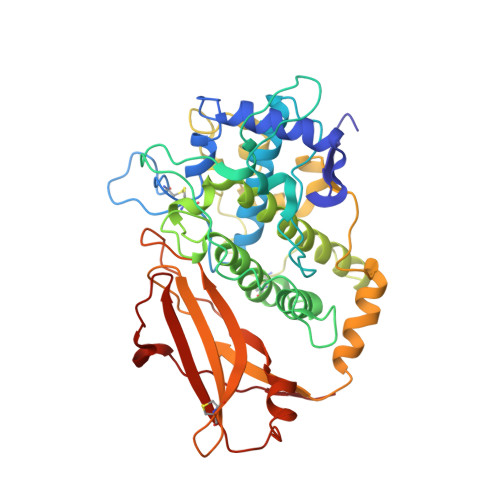
    Structure-function relationships in a molluscan hemocyanin have been investigated by determining the crystal structure of the Rapana thomasiana (gastropod) hemocyanin functional unit RtH2e in deoxygenated form at 3.38 A resolution. This is the first X-ray structure of an unit from the wall of the molluscan hemocyanin cylinder. The crystal structure of RtH2e demonstrates molecular self-assembly of six identical molecules forming a regular hexameric cylinder. This suggests how the functional units are ordered in the wall of the native molluscan hemocyanins. The molecular arrangement is stabilized by specific protomer-to-protomer interactions, which are probably typical for the functional units building the wall of the cylinders. A molecular mechanism for cooperative dioxygen binding in molluscan hemocyanins is proposed on the basis of the molecular interactions between the protomers. In particular, the deoxygenated RtH2e structure reveals a tunnel leading from two opposite sides of the molecule to the active site. The tunnel represents a possible entrance pathway for dioxygen molecules. No such tunnels have been observed in the crystal structure of the oxy-Odg, a functional unit from the Octopus dofleini (cephalopod) hemocyanin in oxygenated form.


  • Organizational Affiliation

    Institute of Biochemistry and Molecular Biology I, University Hospital Hamburg-Eppendorf, c/o DESY, Building 22a, Notkestrasse 85, 22603 Hamburg, Germany.


Macromolecules
Find similar proteins by:  (by identity cutoff)  |  3D Structure
Entity ID: 1
MoleculeChains Sequence LengthOrganismDetailsImage
hemocyanin
A, B, C
408Rapana venosaMutation(s): 0 
UniProt
Find proteins for P83040 (Rapana venosa)
Explore P83040 
Go to UniProtKB:  P83040
Entity Groups  
Sequence Clusters30% Identity50% Identity70% Identity90% Identity95% Identity100% Identity
UniProt GroupP83040
Sequence Annotations
Expand
  • Reference Sequence
Small Molecules
Ligands 2 Unique
IDChains Name / Formula / InChI Key2D Diagram3D Interactions
NAG
Query on NAG

Download Ideal Coordinates CCD File 
D [auth A],
K [auth C],
L [auth C]
2-acetamido-2-deoxy-beta-D-glucopyranose
C8 H15 N O6
OVRNDRQMDRJTHS-FMDGEEDCSA-N
CU
Query on CU

Download Ideal Coordinates CCD File 
E [auth A]
F [auth A]
G [auth B]
H [auth B]
I [auth B]
COPPER (II) ION
Cu
JPVYNHNXODAKFH-UHFFFAOYSA-N
Experimental Data & Validation

Experimental Data

  • Method: X-RAY DIFFRACTION
  • Resolution: 3.30 Å
  • R-Value Free:  0.288 (Depositor) 
  • R-Value Work:  0.248 (Depositor), 0.246 (DCC) 
  • R-Value Observed: 0.250 (Depositor) 
Space Group: P 43 21 2
Unit Cell:
Length ( Å )Angle ( ˚ )
a = 105.486α = 90
b = 105.486β = 90
c = 374.988γ = 90
Software Package:
Software NamePurpose
DENZOdata reduction
SCALEPACKdata scaling
AMoREphasing
REFMACrefinement

Structure Validation

View Full Validation Report


Ligand Structure Quality Assessment 

Created with Raphaël 2.3.0Worse 01 BetterLigand structure goodness of fit to experimental dataBest fitted NAGClick on this verticalbar to view details

Entry History 

Deposition Data

Revision History  (Full details and data files)

  • Version 1.0: 2003-06-03
    Type: Initial release
  • Version 1.1: 2008-04-28
    Changes: Version format compliance
  • Version 1.2: 2011-07-13
    Changes: Advisory, Version format compliance
  • Version 1.3: 2018-07-04
    Changes: Advisory, Data collection, Derived calculations
  • Version 1.4: 2019-11-20
    Changes: Advisory, Derived calculations
  • Version 1.5: 2020-07-29
    Type: Remediation
    Reason: Carbohydrate remediation
    Changes: Advisory, Data collection, Derived calculations, Structure summary
  • Version 1.6: 2024-10-16
    Changes: Advisory, Data collection, Database references, Refinement description, Structure summary