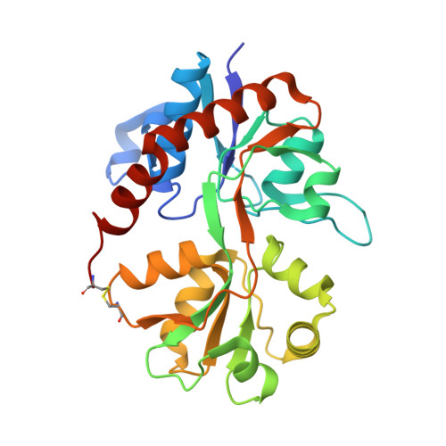
1NNK | pdb_00001nnk

X-ray structure of the GluR2 ligand-binding core (S1S2J) in complex with (S)-ATPA at 1.85 A resolution. Crystallization with zinc ions.


Experimental Data Snapshot

  • Method: X-RAY DIFFRACTION
  • Resolution: 1.85 Å
  • R-Value Free: 
    0.245 (Depositor), 0.240 (DCC) 
  • R-Value Work: 
    0.207 (Depositor), 0.200 (DCC) 

Starting Model: other
View more details

wwPDB Validation   3D Report Full Report


Ligand Structure Quality Assessment 

Created with Raphaël 2.3.0Worse 01 BetterLigand structure goodness of fit to experimental dataBest fitted CE2Click on this verticalbar to view details

This is version 1.5 of the entry. See complete history


Literature

Three-Dimensional Structure of the Ligand-Binding Core of GluR2 in Complex with the Agonist (S)-ATPA: Implications for Receptor Subunit Selectivity.

Lunn, M.L.Hogner, A.Stensbol, T.B.Gouaux, E.Egebjerg, J.Kastrup, J.S.

(2003) J Med Chem 46: 872-875

  • DOI: https://doi.org/10.1021/jm021020+
  • Primary Citation of Related Structures:  
    1NNK, 1NNP

  • PubMed Abstract: 

    Two X-ray structures of the GluR2 ligand-binding core in complex with (S)-2-amino-3-(5-tert-butyl-3-hydroxy-4-isoxazolyl)propionic acid ((S)-ATPA) have been determined with and without Zn(2+) ions. (S)-ATPA induces a domain closure of ca. 21 degrees compared to the apo form. The tert-butyl moiety of (S)-ATPA is buried in a partially hydrophobic pocket and forces the ligand into the glutamate-like binding mode. The structures provide new insight into the molecular basis of agonist selectivity between AMPA and kainate receptors.


  • Organizational Affiliation

    Department of Medicinal Chemistry, Pharmaceutical University of Denmark, Universitetsparken 2, DK-2100 Copenhagen, Denmark.


Macromolecules
Find similar proteins by:  (by identity cutoff)  |  3D Structure
Entity ID: 1
MoleculeChains Sequence LengthOrganismDetailsImage
Glutamate receptor 2263Rattus norvegicusMutation(s): 0 
UniProt
Find proteins for P19491 (Rattus norvegicus)
Explore P19491 
Go to UniProtKB:  P19491
Entity Groups  
Sequence Clusters30% Identity50% Identity70% Identity90% Identity95% Identity100% Identity
UniProt GroupP19491
Sequence Annotations
Expand
  • Reference Sequence
Experimental Data & Validation

Experimental Data

  • Method: X-RAY DIFFRACTION
  • Resolution: 1.85 Å
  • R-Value Free:  0.245 (Depositor), 0.240 (DCC) 
  • R-Value Work:  0.207 (Depositor), 0.200 (DCC) 
Space Group: P 21 21 2
Unit Cell:
Length ( Å )Angle ( ˚ )
a = 54.258α = 90
b = 111.229β = 90
c = 46.643γ = 90
Software Package:
Software NamePurpose
DENZOdata reduction
SCALEPACKdata scaling
AMoREphasing
CNSrefinement

Structure Validation

View Full Validation Report


Ligand Structure Quality Assessment 

Created with Raphaël 2.3.0Worse 01 BetterLigand structure goodness of fit to experimental dataBest fitted CE2Click on this verticalbar to view details

Entry History 

Deposition Data

Revision History  (Full details and data files)

  • Version 1.0: 2003-03-04
    Type: Initial release
  • Version 1.1: 2008-04-29
    Changes: Version format compliance
  • Version 1.2: 2011-07-13
    Changes: Derived calculations, Version format compliance
  • Version 1.3: 2017-08-16
    Changes: Data collection, Refinement description, Source and taxonomy
  • Version 1.4: 2024-04-03
    Changes: Data collection, Database references, Derived calculations, Refinement description, Structure summary
  • Version 1.5: 2024-10-16
    Changes: Structure summary