2AVW | pdb_00002avw

Crystal structure of monoclinic form of streptococcus Mac-1


Experimental Data Snapshot

  • Method: X-RAY DIFFRACTION
  • Resolution: 2.00 Å
  • R-Value Free: 
    0.267 (Depositor) 
  • R-Value Work: 
    0.218 (Depositor), 0.220 (DCC) 
  • R-Value Observed: 
    0.220 (Depositor) 

wwPDB Validation   3D Report Full Report


This is version 1.4 of the entry. See complete history


Literature

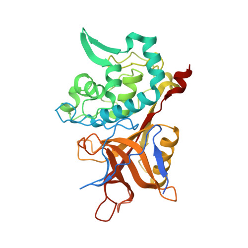
Crystal structure of group a streptococcus mac-1: insight into dimer-mediated specificity for recognition of human IgG.

Agniswamy, J.Nagiec, M.J.Liu, M.Schuck, P.Musser, J.M.Sun, P.D.

(2006) Structure 14: 225-235

  • DOI: https://doi.org/10.1016/j.str.2005.10.012
  • Primary Citation of Related Structures:  
    2AU1, 2AVW

  • PubMed Abstract: 

    Group A Streptococcus secretes cysteine proteases named Mac-1 and Mac-2 that mediate host immune evasion by targeting both IgG and Fc receptors. Here, we report the crystal structures of Mac-1 and its catalytically inactive C94A mutant in two different crystal forms. Despite the lack of sequence homology, Mac-1 adopts the canonical papain fold. Alanine mutations at the active site confirmed the critical residues involved in a papain-like catalytic mechanism. Mac-1 forms a symmetric dimer in both crystal forms and displays the unique dimer interface among papain superfamily members. Mutations at the dimer interface resulted in a significant reduction in IgG binding and catalysis, suggesting that the dimer contributes to both IgG specificity and enzyme cooperativity. A tunnel observed at the dimer interface constitutes a target for designing potential Mac-1-specific antimicrobial agents. The structures also offer insight into the functional difference between Mac-1 and Mac-2.


  • Organizational Affiliation
    • Structural Immunology Section, Laboratory of Immunogenetics, National Institute of Allergy and Infectious Diseases, National Institutes of Health, 12441 Parklawn Drive, Rockville, Maryland 20852, USA.

Macromolecules
Find similar proteins by:  (by identity cutoff)  |  3D Structure
Entity ID: 1
MoleculeChains Sequence LengthOrganismDetailsImage
IgG-degrading protease
A, B, C, D, E
A, B, C, D, E, F
311Streptococcus pyogenes MGAS5005Mutation(s): 1 
Gene Names: mac
UniProt
Find proteins for Q7DAM2 (Streptococcus pyogenes serotype M1)
Explore Q7DAM2 
Go to UniProtKB:  Q7DAM2
Entity Groups  
Sequence Clusters30% Identity50% Identity70% Identity90% Identity95% Identity100% Identity
UniProt GroupQ7DAM2
Sequence Annotations
Expand
  • Reference Sequence
Experimental Data & Validation

Experimental Data

  • Method: X-RAY DIFFRACTION
  • Resolution: 2.00 Å
  • R-Value Free:  0.267 (Depositor) 
  • R-Value Work:  0.218 (Depositor), 0.220 (DCC) 
  • R-Value Observed: 0.220 (Depositor) 
Space Group: P 1 21 1
Unit Cell:
Length ( Å )Angle ( ˚ )
a = 106.986α = 90
b = 65.464β = 105.81
c = 137.244γ = 90
Software Package:
Software NamePurpose
HKL-2000data collection
SCALEPACKdata scaling
CNSrefinement
HKL-2000data reduction
CNSphasing

Structure Validation

View Full Validation Report


Entry History 

Deposition Data

Revision History  (Full details and data files)

  • Version 1.0: 2006-02-28
    Type: Initial release
  • Version 1.1: 2008-05-01
    Changes: Version format compliance
  • Version 1.2: 2011-07-13
    Changes: Source and taxonomy, Version format compliance
  • Version 1.3: 2021-10-20
    Changes: Database references, Derived calculations
  • Version 1.4: 2024-02-14
    Changes: Data collection