


Crystal Structure of Factor VIIa/soluble Tissue Factor complexed with Glu-Gly-Arg-Chloromethyl ketone
Bajaj, S.P., Schmidt, A.E., Padmanabhan, K.To be published.
Experimental Data Snapshot
Entity ID: 1 | |||||
|---|---|---|---|---|---|
| Molecule | Chains | Sequence Length | Organism | Details | Image |
| Coagulation factor VII light chain | A [auth L] | 142 | Homo sapiens | Mutation(s): 0 Gene Names: F7 EC: 3.4.21.21 | 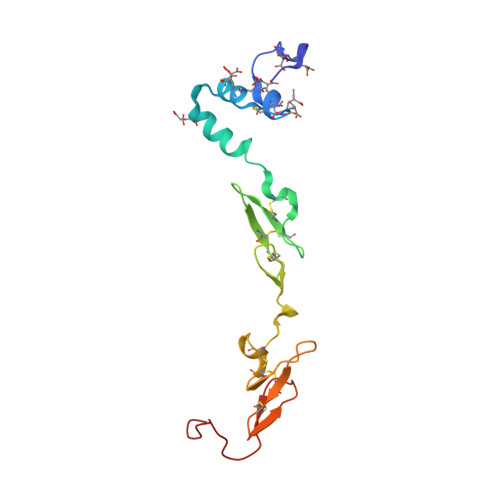 |
UniProt & NIH Common Fund Data Resources | |||||
Find proteins for P08709 (Homo sapiens) Explore P08709 Go to UniProtKB: P08709 | |||||
PHAROS: P08709 GTEx: ENSG00000057593 | |||||
Entity Groups | |||||
| Sequence Clusters | 30% Identity50% Identity70% Identity90% Identity95% Identity100% Identity | ||||
| UniProt Group | P08709 | ||||
Glycosylation | |||||
| Glycosylation Sites: 2 | Go to GlyGen: P08709-1 | ||||
Sequence AnnotationsExpand | |||||
| |||||
Entity ID: 2 | |||||
|---|---|---|---|---|---|
| Molecule | Chains | Sequence Length | Organism | Details | Image |
| Coagulation factor VII heavy chain | B [auth H] | 254 | Homo sapiens | Mutation(s): 0 Gene Names: F7 EC: 3.4.21.21 | 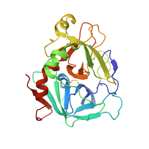 |
UniProt & NIH Common Fund Data Resources | |||||
Find proteins for P08709 (Homo sapiens) Explore P08709 Go to UniProtKB: P08709 | |||||
PHAROS: P08709 GTEx: ENSG00000057593 | |||||
Entity Groups | |||||
| Sequence Clusters | 30% Identity50% Identity70% Identity90% Identity95% Identity100% Identity | ||||
| UniProt Group | P08709 | ||||
Sequence AnnotationsExpand | |||||
| |||||
Entity ID: 3 | |||||
|---|---|---|---|---|---|
| Molecule | Chains | Sequence Length | Organism | Details | Image |
| Tissue factor | C [auth T] | 205 | Homo sapiens | Mutation(s): 0 Gene Names: F3 | 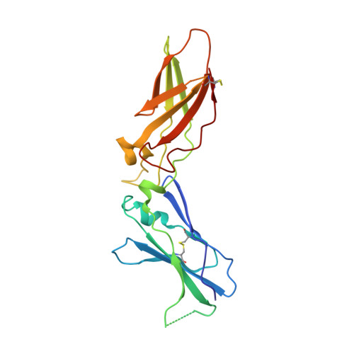 |
UniProt & NIH Common Fund Data Resources | |||||
Find proteins for P13726 (Homo sapiens) Explore P13726 Go to UniProtKB: P13726 | |||||
PHAROS: P13726 GTEx: ENSG00000117525 | |||||
Entity Groups | |||||
| Sequence Clusters | 30% Identity50% Identity70% Identity90% Identity95% Identity100% Identity | ||||
| UniProt Group | P13726 | ||||
Sequence AnnotationsExpand | |||||
| |||||
| Ligands 8 Unique | |||||
|---|---|---|---|---|---|
| ID | Chains | Name / Formula / InChI Key | 2D Diagram | 3D Interactions | |
| 0GJ Query on 0GJ | M [auth H] | L-alpha-glutamyl-N-{(1S)-4-{[amino(iminio)methyl]amino}-1-[(1S)-2-chloro-1-hydroxyethyl]butyl}glycinamide C14 H28 Cl N6 O5 XELWNHKFCNMWQO-LPEHRKFASA-O |  | ||
| GLC Query on GLC | D [auth L] | alpha-D-glucopyranose C6 H12 O6 WQZGKKKJIJFFOK-DVKNGEFBSA-N |  | ||
| FUC Query on FUC | E [auth L] | alpha-L-fucopyranose C6 H12 O5 SHZGCJCMOBCMKK-SXUWKVJYSA-N |  | ||
| ZN Query on ZN | P [auth H], Q [auth H] | ZINC ION Zn PTFCDOFLOPIGGS-UHFFFAOYSA-N |  | ||
| CA Query on CA | F [auth L] G [auth L] I [auth L] J [auth L] L | CALCIUM ION Ca BHPQYMZQTOCNFJ-UHFFFAOYSA-N |  | ||
| CL Query on CL | R [auth H], S [auth H], T | CHLORIDE ION Cl VEXZGXHMUGYJMC-UHFFFAOYSA-M |  | ||
| MG Query on MG | H [auth L], K [auth L] | MAGNESIUM ION Mg JLVVSXFLKOJNIY-UHFFFAOYSA-N |  | ||
| NA Query on NA | O [auth H] | SODIUM ION Na FKNQFGJONOIPTF-UHFFFAOYSA-N |  | ||
| Modified Residues 1 Unique | |||||
|---|---|---|---|---|---|
| ID | Chains | Type | Formula | 2D Diagram | Parent |
| CGU Query on CGU | A [auth L] | L-PEPTIDE LINKING | C6 H9 N O6 |  | GLU |
Entity ID: 8 | |||||
|---|---|---|---|---|---|
| ID | Chains | Name | Type/Class | 2D Diagram | 3D Interactions |
| PRD_000288 (0GJ) Query on PRD_000288 | M [auth H] | GLU-GLY-ARG-CHLOROMETHYL KETONE | Peptide-like / Inhibitor | | |
| Length ( Å ) | Angle ( ˚ ) |
|---|---|
| a = 70.28 | α = 90 |
| b = 81.11 | β = 90 |
| c = 126.86 | γ = 90 |
| Software Name | Purpose |
|---|---|
| X-PLOR | model building |
| X-PLOR | refinement |
| DENZO | data reduction |
| SCALEPACK | data scaling |
| X-PLOR | phasing |


