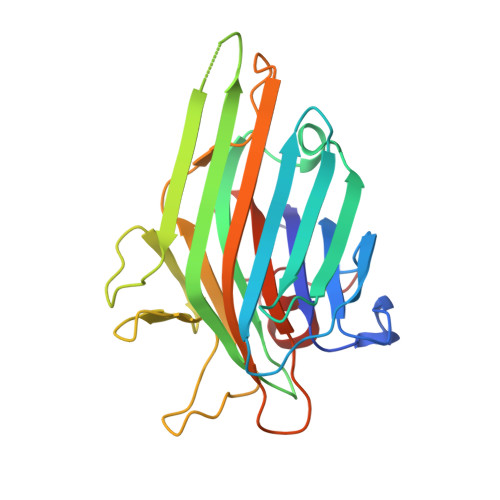
2EF6

Canavalia gladiata lectin complexed with Man1-3Man-OMe


Experimental Data Snapshot

  • Method: X-RAY DIFFRACTION
  • Resolution: 2.10 Å
  • R-Value Free: 0.245 
  • R-Value Work: 0.189 
  • R-Value Observed: 0.192 

Starting Model: experimental
View more details

wwPDB Validation   3D Report Full Report


This is version 2.1 of the entry. See complete history


Literature

Structural analysis of Canavalia maritima and Canavalia gladiata lectins complexed with different dimannosides: New insights into the understanding of the structure-biological activity relationship in legume lectins

Bezerra, G.A.Oliveira, T.M.Moreno, F.B.M.B.de Souza, E.P.da Rocha, B.A.M.Benevides, R.G.Delatorre, P.de Azevedo Jr., W.F.Cavada, B.S.

(2007) J Struct Biol 160: 168-176

  • DOI: https://doi.org/10.1016/j.jsb.2007.07.012
  • Primary Citation of Related Structures:  
    2EF6, 2OVU, 2OW4, 2P2K, 2P34, 2P37

  • PubMed Abstract: 

    Plant lectins, especially those purified from species of the Leguminosae family, represent the best studied group of carbohydrate-binding proteins. The legume lectins from Diocleinae subtribe are highly similar proteins that present significant differences in the potency/efficacy of their biological activities. The structural studies of the interactions between lectins and sugars may clarify the origin of the distinct biological activities observed in this high similar class of proteins. In this way, this work presents a crystallographic study of the ConM and CGL (agglutinins from Canavalia maritima and Canavalia gladiata, respectively) in the following complexes: ConM/CGL:Man(alpha1-2)Man(alpha1-O)Me, ConM/CGL:Man(alpha1-3)Man(alpha1-O)Me and ConM/CGL:Man(alpha1-4)Man(alpha1-O)Me, which crystallized in different conditions and space group from the native proteins. The structures were solved by molecular replacement, presenting satisfactory values for R(factor) and R(free). Comparisons between ConM, CGL and ConA (Canavalia ensiformis lectin) binding mode with the dimannosides in subject, presented different interactions patterns, which may account for a structural explanation of the distincts biological properties observed in the lectins of Diocleinae subtribe.


  • Organizational Affiliation

    Departamento de Bioquímica e Biologia Molecular, Universidade Federal do Ceará, Biomol-LAB, Campus do Pici S/N, Fortaleza, Ceará, Brazil.


Macromolecules
Find similar proteins by:  (by identity cutoff)  |  3D Structure
Entity ID: 1
MoleculeChains Sequence LengthOrganismDetailsImage
Concanavalin A
A, B, C, D
237Canavalia gladiataMutation(s): 0 
UniProt
Find proteins for P14894 (Canavalia gladiata)
Explore P14894 
Go to UniProtKB:  P14894
Entity Groups  
Sequence Clusters30% Identity50% Identity70% Identity90% Identity95% Identity100% Identity
UniProt GroupP14894
Sequence Annotations
Expand
  • Reference Sequence
Oligosaccharides

Help

Entity ID: 2
MoleculeChains Length2D Diagram Glycosylation3D Interactions
alpha-D-mannopyranose-(1-3)-methyl alpha-D-mannopyranoside
E, F, G, H
2N/A
Glycosylation Resources
GlyTouCan:  G59594NN
GlyCosmos:  G59594NN
Small Molecules
Ligands 2 Unique
IDChains Name / Formula / InChI Key2D Diagram3D Interactions
MN
Query on MN

Download Ideal Coordinates CCD File 
J [auth A],
L [auth B],
N [auth C],
P [auth D]
MANGANESE (II) ION
Mn
WAEMQWOKJMHJLA-UHFFFAOYSA-N
CA
Query on CA

Download Ideal Coordinates CCD File 
I [auth A],
K [auth B],
M [auth C],
O [auth D]
CALCIUM ION
Ca
BHPQYMZQTOCNFJ-UHFFFAOYSA-N
Experimental Data & Validation

Experimental Data

  • Method: X-RAY DIFFRACTION
  • Resolution: 2.10 Å
  • R-Value Free: 0.245 
  • R-Value Work: 0.189 
  • R-Value Observed: 0.192 
  • Space Group: P 32
Unit Cell:
Length ( Å )Angle ( ˚ )
a = 69.391α = 90
b = 69.391β = 90
c = 161.298γ = 120
Software Package:
Software NamePurpose
REFMACrefinement
MARCCDdata collection
MOSFLMdata reduction
SCALAdata scaling
MOLREPphasing

Structure Validation

View Full Validation Report


Entry History 

Revision History  (Full details and data files)

  • Version 1.0: 2007-10-30
    Type: Initial release
  • Version 1.1: 2011-07-13
    Changes: Version format compliance
  • Version 1.2: 2017-10-11
    Changes: Advisory, Database references, Source and taxonomy
  • Version 2.0: 2020-07-29
    Type: Remediation
    Reason: Carbohydrate remediation
    Changes: Advisory, Atomic model, Data collection, Derived calculations, Non-polymer description, Structure summary
  • Version 2.1: 2023-10-25
    Changes: Data collection, Database references, Refinement description, Structure summary