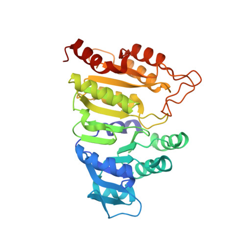
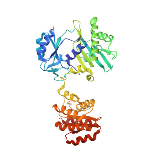
2FPG | pdb_00002fpg

Crystal structure of pig GTP-specific succinyl-CoA synthetase in complex with GDP


Experimental Data Snapshot

  • Method: X-RAY DIFFRACTION
  • Resolution: 2.96 Å
  • R-Value Free: 
    0.275 (Depositor), 0.270 (DCC) 
  • R-Value Work: 
    0.201 (Depositor), 0.200 (DCC) 
  • R-Value Observed: 
    0.205 (Depositor) 

wwPDB Validation   3D Report Full Report


Ligand Structure Quality Assessment 

Created with Raphaël 2.3.0Worse 01 BetterLigand structure goodness of fit to experimental dataBest fitted GDPClick on this verticalbar to view details

This is version 1.4 of the entry. See complete history


Literature

Interactions of GTP with the ATP-grasp Domain of GTP-specific Succinyl-CoA Synthetase

Fraser, M.E.Hayakawa, K.Hume, M.S.Ryan, D.G.Brownie, E.R.

(2006) J Biological Chem 281: 11058-11065

  • DOI: https://doi.org/10.1074/jbc.M511785200
  • Primary Citation of Related Structures:  
    2FP4, 2FPG, 2FPI, 2FPP

  • PubMed Abstract: 

    Two isoforms of succinyl-CoA synthetase exist in mammals, one specific for ATP and the other for GTP. The GTP-specific form of pig succinyl-CoA synthetase has been crystallized in the presence of GTP and the structure determined to 2.1 A resolution. GTP is bound in the ATP-grasp domain, where interactions of the guanine base with a glutamine residue (Gln-20beta) and with backbone atoms provide the specificity. The gamma-phosphate interacts with the side chain of an arginine residue (Arg-54beta) and with backbone amide nitrogen atoms, leading to tight interactions between the gamma-phosphate and the protein. This contrasts with the structures of ATP bound to other members of the family of ATP-grasp proteins where the gamma-phosphate is exposed, free to react with the other substrate. To test if GDP would interact with GTP-specific succinyl-CoA synthetase in the same way that ADP interacts with other members of the family of ATP-grasp proteins, the structure of GDP bound to GTP-specific succinyl-CoA synthetase was also determined. A comparison of the conformations of GTP and GDP shows that the bases adopt the same position but that changes in conformation of the ribose moieties and the alpha- and beta-phosphates allow the gamma-phosphate to interact with the arginine residue and amide nitrogen atoms in GTP, while the beta-phosphate interacts with these residues in GDP. The complex of GTP with succinyl-CoA synthetase shows that the enzyme is able to protect GTP from hydrolysis when the active-site histidine residue is not in position to be phosphorylated.


  • Organizational Affiliation

    Department of Biological Sciences, University of Calgary, Calgary, Alberta T2N 1N4, Canada. frasm@ucalgary.ca


Macromolecules
Find similar proteins by:  (by identity cutoff)  |  3D Structure
Entity ID: 1
MoleculeChains Sequence LengthOrganismDetailsImage
Succinyl-CoA ligase [GDP-forming] alpha-chain, mitochondrial305Sus scrofaMutation(s): 0 
Gene Names: SUCLG1
EC: 6.2.1.4 (PDB Primary Data), 6.2.1.5 (UniProt)
UniProt
Find proteins for O19069 (Sus scrofa)
Explore O19069 
Go to UniProtKB:  O19069
Entity Groups  
Sequence Clusters30% Identity50% Identity70% Identity90% Identity95% Identity100% Identity
UniProt GroupO19069
Sequence Annotations
Expand
  • Reference Sequence
Find similar proteins by:  (by identity cutoff)  |  3D Structure
Entity ID: 2
MoleculeChains Sequence LengthOrganismDetailsImage
Succinyl-CoA ligase [GDP-forming] beta-chain, mitochondrial395Sus scrofaMutation(s): 0 
Gene Names: SUCLG2
EC: 6.2.1.4
UniProt
Find proteins for P53590 (Sus scrofa)
Explore P53590 
Go to UniProtKB:  P53590
Entity Groups  
Sequence Clusters30% Identity50% Identity70% Identity90% Identity95% Identity100% Identity
UniProt GroupP53590
Sequence Annotations
Expand
  • Reference Sequence
Experimental Data & Validation

Experimental Data

  • Method: X-RAY DIFFRACTION
  • Resolution: 2.96 Å
  • R-Value Free:  0.275 (Depositor), 0.270 (DCC) 
  • R-Value Work:  0.201 (Depositor), 0.200 (DCC) 
  • R-Value Observed: 0.205 (Depositor) 
Space Group: P 65
Unit Cell:
Length ( Å )Angle ( ˚ )
a = 133.78α = 90
b = 133.78β = 90
c = 73.09γ = 120
Software Package:
Software NamePurpose
SCALAdata scaling
CNSrefinement
PDB_EXTRACTdata extraction
MOSFLMdata reduction
CCP4data scaling
CNSphasing

Structure Validation

View Full Validation Report


Ligand Structure Quality Assessment 

Created with Raphaël 2.3.0Worse 01 BetterLigand structure goodness of fit to experimental dataBest fitted GDPClick on this verticalbar to view details

Entry History 

Deposition Data

  • Released Date: 2006-02-21 
  • Deposition Author(s): Fraser, M.E.

Revision History  (Full details and data files)

  • Version 1.0: 2006-02-21
    Type: Initial release
  • Version 1.1: 2008-05-01
    Changes: Version format compliance
  • Version 1.2: 2011-07-13
    Changes: Version format compliance
  • Version 1.3: 2017-10-18
    Changes: Refinement description
  • Version 1.4: 2024-02-14
    Changes: Data collection, Database references, Derived calculations