NMR structural studies of the first catalytic half-domain of ubiquitin activating enzyme.
Jaremko, M., Jaremko, L., Nowakowski, M., Wojciechowski, M., Szczepanowski, R.H., Panecka, R., Zhukov, I., Bochtler, M., Ejchart, A.(2014) J Struct Biol 185: 69-78
- PubMed: 24211821
- DOI: https://doi.org/10.1016/j.jsb.2013.10.020
- Primary Citation of Related Structures:
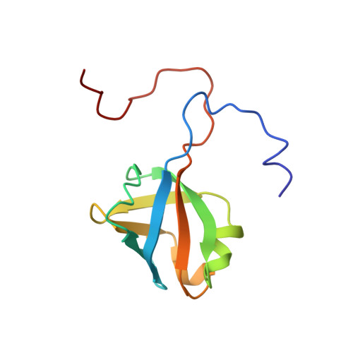
2LZJ - PubMed Abstract:
We report a high resolution NMR structure and (15)N relaxation studies of the first catalytic cysteine half-domain (FCCH) of the mouse ubiquitin-activating enzyme E1, together with interaction studies of FCCH and the other catalytic E1 subdomain - SCCH (second catalytic cysteine half-domain). In solution, mouse FCCH forms a well-defined six-stranded antiparallel β-barrel structure, a common fold for many proteins with a variety of cellular functions. (15)N relaxation data reveal FCCH complex backbone dynamics and indicate which residues experience slow intramolecular motions. Some of these residues make contacts with the polar face of ubiquitin in the co-crystal structure of yeast E1 and ubiquitin. However, the titration of FCCH with ubiquitin does not show any visible chemical shift changes in the 2D (1)H/(15)N HSQC spectra of the FCCH. The 2D (1)H/(15)N HSQC experiments performed both for each catalytic half-domain individually and for their equimolar mixture in the milimolar concentration range display no detectable chemical shift perturbation, suggesting a lack of interaction between the two subdomains unless they are covalently linked via the adenylation domain.
- Max Planck Institute for Biophysical Chemistry, Department for NMR-based Structural Biology, Am Fassber 11, 37077 Göttingen, Germany.
Organizational Affiliation: