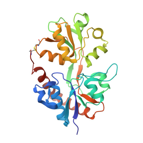
2P2A | pdb_00002p2a

X-ray structure of the GluR2 ligand binding core (S1S2J) in complex with 2-Bn-tet-AMPA at 2.26A resolution


Experimental Data Snapshot

  • Method: X-RAY DIFFRACTION
  • Resolution: 2.26 Å
  • R-Value Free: 
    0.273 (Depositor), 0.260 (DCC) 
  • R-Value Work: 
    0.211 (Depositor), 0.200 (DCC) 

Starting Model: experimental
View more details

wwPDB Validation   3D Report Full Report


Ligand Structure Quality Assessment 

Created with Raphaël 2.3.0Worse 01 BetterLigand structure goodness of fit to experimental dataBest fitted MP9Click on this verticalbar to view details

This is version 1.6 of the entry. See complete history


Literature

A tetrazolyl-substituted subtype-selective AMPA receptor agonist.

Vogensen, S.B.Frydenvang, K.Greenwood, J.R.Postorino, G.Nielsen, B.Pickering, D.S.Ebert, B.Bolcho, U.Egebjerg, J.Gajhede, M.Kastrup, J.S.Johansen, T.N.Clausen, R.P.Krogsgaard-Larsen, P.

(2007) J Med Chem 50: 2408-2414

  • DOI: https://doi.org/10.1021/jm061439q
  • Primary Citation of Related Structures:  
    2P2A

  • PubMed Abstract: 

    Replacement of the methyl group of the AMPA receptor agonist 2-amino-3-[3-hydroxy-5-(2-methyl-2H-5-tetrazolyl)-4-isoxazolyl]propionic acid (2-Me-Tet-AMPA) with a benzyl group provided the first AMPA receptor agonist, compound 7, capable of discriminating GluR2-4 from GluR1 by its more than 10-fold preference for the former receptor subtypes. An X-ray crystallographic analysis of this new analogue in complex with the GluR2-S1S2J construct shows that accommodation of the benzyl group creates a previously unobserved pocket in the receptor, which may explain the remarkable pharmacological profile of compound 7.


  • Organizational Affiliation

    Department of Medicinal Chemistry, The Faculty of Pharmaceutical Sciences, University of Copenhagen, 2 Universitetsparken, DK-2100 Copenhagen, Denmark.


Macromolecules
Find similar proteins by:  (by identity cutoff)  |  3D Structure
Entity ID: 1
MoleculeChains Sequence LengthOrganismDetailsImage
Glutamate receptor 2
A, B
263Rattus norvegicusMutation(s): 0 
Gene Names: Gria2
UniProt
Find proteins for P19491 (Rattus norvegicus)
Explore P19491 
Go to UniProtKB:  P19491
Entity Groups  
Sequence Clusters30% Identity50% Identity70% Identity90% Identity95% Identity100% Identity
UniProt GroupP19491
Sequence Annotations
Expand
  • Reference Sequence
Experimental Data & Validation

Experimental Data

  • Method: X-RAY DIFFRACTION
  • Resolution: 2.26 Å
  • R-Value Free:  0.273 (Depositor), 0.260 (DCC) 
  • R-Value Work:  0.211 (Depositor), 0.200 (DCC) 
Space Group: P 21 21 2
Unit Cell:
Length ( Å )Angle ( ˚ )
a = 98.12α = 90
b = 121.98β = 90
c = 47.2γ = 90
Software Package:
Software NamePurpose
CNSrefinement
PDB_EXTRACTdata extraction
MOSFLMdata reduction
SCALAdata scaling
AMoREphasing

Structure Validation

View Full Validation Report


Ligand Structure Quality Assessment 

Created with Raphaël 2.3.0Worse 01 BetterLigand structure goodness of fit to experimental dataBest fitted MP9Click on this verticalbar to view details

Entry History 

Deposition Data

Revision History  (Full details and data files)

  • Version 1.0: 2007-06-19
    Type: Initial release
  • Version 1.1: 2008-05-01
    Changes: Version format compliance
  • Version 1.2: 2011-07-13
    Changes: Version format compliance
  • Version 1.3: 2017-08-16
    Changes: Data collection, Refinement description, Source and taxonomy
  • Version 1.4: 2022-12-21
    Changes: Database references, Derived calculations
  • Version 1.5: 2023-09-20
    Changes: Data collection, Refinement description
  • Version 1.6: 2024-11-20
    Changes: Structure summary