2YEX

Synthesis and evaluation of triazolones as checkpoint kinase 1 inhibitors


Experimental Data Snapshot

  • Method: X-RAY DIFFRACTION
  • Resolution: 1.30 Å
  • R-Value Free: 
    0.164 (Depositor), 0.180 (DCC) 
  • R-Value Work: 
    0.134 (Depositor), 0.150 (DCC) 
  • R-Value Observed: 
    0.135 (Depositor) 

wwPDB Validation   3D Report Full Report


Ligand Structure Quality Assessment 


This is version 1.1 of the entry. See complete history


Literature

Synthesis and Evaluation of Triazolones as Checkpoint Kinase 1 Inhibitors.

Oza, V.Ashwell, S.Brassil, P.Breed, J.Ezhuthachan, J.Deng, C.Grondine, M.Horn, C.Liu, D.Lyne, P.Newcombe, N.Pass, M.Read, J.Su, M.Toader, D.Yu, D.Yu, Y.Zabludoff, S.

(2012) Bioorg Med Chem Lett 22: 2330

  • DOI: https://doi.org/10.1016/j.bmcl.2012.01.043
  • Primary Citation of Related Structures:  
    2YER, 2YEX

  • PubMed Abstract: 

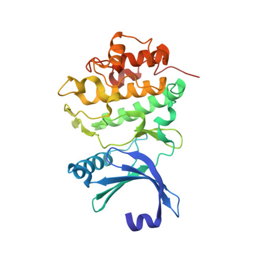
    Checkpoint kinase 1 (Chk1, CHEK1) is a Ser/Thr protein kinase that plays a key role in mediating the cellular response to DNA-damage. Synthesis and evaluation of a previously described class of Chk1 inhibitors, triazoloquinolones/triazolones (TZs) is further described herein. Our investigation of structure-activity relationships led to the identification of potent inhibitors 14c, 14h and 16e. Key challenges included modulation of physicochemical properties and pharmacokinetic (PK) parameters to enable compound testing in a Chk1 specific hollow fiber pharmacodynamic model. In this model, 16e was shown to abrogate topotecan-induced cell cycle arrest in a dose dependent manner. The demonstrated activity of TZs in this model in combination with a chemotherapeutic agent as well as radiotherapy validates this series of Chk1 inhibitors. X-ray crystal structures (PDB code: 2YEX and 2YER) for an initial lead and an optimized analog are also presented.


  • Organizational Affiliation

    AstraZeneca R&D Boston, 35 Gatehouse Drive, Waltham, MA 02451, USA. Vibha.Oza@astrazeneca.com


Macromolecules
Find similar proteins by:  (by identity cutoff)  |  3D Structure
Entity ID: 1
MoleculeChains Sequence LengthOrganismDetailsImage
SERINE/THREONINE-PROTEIN KINASE CHK1276Homo sapiensMutation(s): 0 
EC: 2.7.11.1
UniProt & NIH Common Fund Data Resources
Find proteins for O14757 (Homo sapiens)
Explore O14757 
Go to UniProtKB:  O14757
PHAROS:  O14757
GTEx:  ENSG00000149554 
Entity Groups  
Sequence Clusters30% Identity50% Identity70% Identity90% Identity95% Identity100% Identity
UniProt GroupO14757
Sequence Annotations
Expand
  • Reference Sequence
Small Molecules
Ligands 3 Unique
IDChains Name / Formula / InChI Key2D Diagram3D Interactions
YEX
Query on YEX

Download Ideal Coordinates CCD File 
I [auth A]5-METHYL-8-(1H-PYRROL-2-YL)[1,2,4]TRIAZOLO[4,3-A]QUINOLIN-1(2H)-ONE
C15 H12 N4 O
XHMYMTXQNURDOQ-UHFFFAOYSA-N
SO4
Query on SO4

Download Ideal Coordinates CCD File 
J [auth A]SULFATE ION
O4 S
QAOWNCQODCNURD-UHFFFAOYSA-L
GOL
Query on GOL

Download Ideal Coordinates CCD File 
B [auth A]
C [auth A]
D [auth A]
E [auth A]
F [auth A]
B [auth A],
C [auth A],
D [auth A],
E [auth A],
F [auth A],
G [auth A],
H [auth A]
GLYCEROL
C3 H8 O3
PEDCQBHIVMGVHV-UHFFFAOYSA-N
Binding Affinity Annotations 
IDSourceBinding Affinity
YEX BindingDB:  2YEX IC50: 0.1 (nM) from 1 assay(s)
Experimental Data & Validation

Experimental Data

  • Method: X-RAY DIFFRACTION
  • Resolution: 1.30 Å
  • R-Value Free:  0.164 (Depositor), 0.180 (DCC) 
  • R-Value Work:  0.134 (Depositor), 0.150 (DCC) 
  • R-Value Observed: 0.135 (Depositor) 
Space Group: P 21 21 21
Unit Cell:
Length ( Å )Angle ( ˚ )
a = 41.352α = 90
b = 70.77β = 90
c = 104.327γ = 90
Software Package:
Software NamePurpose
REFMACrefinement
MOSFLMdata reduction
SCALAdata scaling
AMoREphasing

Structure Validation

View Full Validation Report


Ligand Structure Quality Assessment 


Entry History 

Deposition Data

Revision History  (Full details and data files)

  • Version 1.0: 2012-03-14
    Type: Initial release
  • Version 1.1: 2024-05-08
    Changes: Data collection, Database references, Derived calculations, Other