2Z9X | pdb_00002z9x

Crystal structure of pyridoxamine-pyruvate aminotransferase complexed with pyridoxyl-L-alanine


Experimental Data Snapshot

  • Method: X-RAY DIFFRACTION
  • Resolution: 1.94 Å
  • R-Value Free: 
    0.184 (Depositor), 0.190 (DCC) 
  • R-Value Work: 
    0.145 (Depositor), 0.150 (DCC) 
  • R-Value Observed: 
    0.147 (Depositor) 

Starting Model: experimental
View more details

wwPDB Validation   3D Report Full Report


Ligand Structure Quality Assessment 

Created with Raphaël 2.3.0Worse 01 BetterLigand structure goodness of fit to experimental dataBest fitted PXLClick on this verticalbar to view details

This is version 1.2 of the entry. See complete history


Literature

Crystal structure of pyridoxamine-pyruvate aminotransferase from Mesorhizobium loti MAFF303099

Yoshikane, Y.Yokochi, N.Yamasaki, M.Mizutani, K.Ohnishi, K.Mikami, B.Hayashi, H.Yagi, T.

(2008) J Biological Chem 283: 1120-1127

  • DOI: https://doi.org/10.1074/jbc.M708061200
  • Primary Citation of Related Structures:  
    2Z9U, 2Z9V, 2Z9W, 2Z9X

  • PubMed Abstract: 

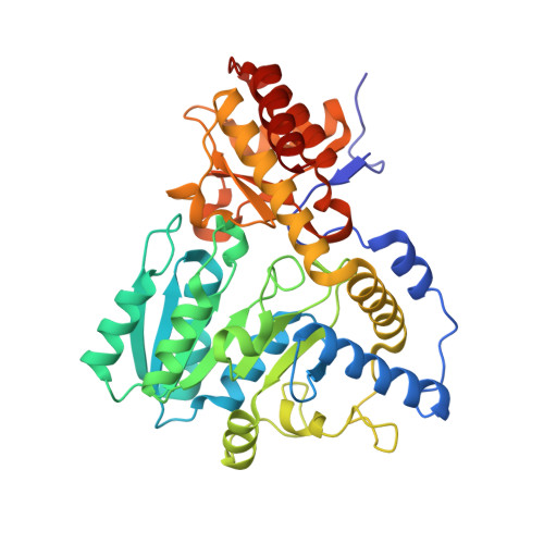
    Pyridoxamine-pyruvate aminotransferase (PPAT; EC 2.6.1.30) is a pyridoxal 5'-phosphate-independent aminotransferase and catalyzes reversible transamination between pyridoxamine and pyruvate to form pyridoxal and L-alanine. The crystal structure of PPAT from Mesorhizobium loti has been solved in space group P4(3)2(1)2 and was refined to an R factor of 15.6% (R(free) = 20.6%) at 2.0 A resolution. In addition, the structures of PPAT in complexes with pyridoxamine, pyridoxal, and pyridoxyl-L-alanine have been refined to R factors of 15.6, 15.4, and 14.5% (R(free) = 18.6, 18.1, and 18.4%) at 1.7, 1.7, and 2.0 A resolution, respectively. PPAT is a homotetramer and each subunit is composed of a large N-terminal domain, consisting of seven beta-sheets and eight alpha-helices, and a smaller C-terminal domain, consisting of three beta-sheets and four alpha-helices. The substrate pyridoxal is bound through an aldimine linkage to Lys-197 in the active site. The alpha-carboxylate group of the substrate amino/keto acid is hydrogen-bonded to Arg-336 and Arg-345. The structures revealed that the bulky side chain of Glu-68 interfered with the binding of the phosphate moiety of pyridoxal 5'-phosphate and made PPAT specific to pyridoxal. The reaction mechanism of the enzyme is discussed based on the structures and kinetics results.


  • Organizational Affiliation

    Department of Bioresources Science, Faculty of Agriculture, Kochi University, Monobe-Otsu 200, Nankoku, Kochi 783-8502, Japan.


Macromolecules
Find similar proteins by:  (by identity cutoff)  |  3D Structure
Entity ID: 1
MoleculeChains Sequence LengthOrganismDetailsImage
Aspartate aminotransferase
A, B
392Mesorhizobium lotiMutation(s): 0 
Gene Names: mlr6806
EC: 2.6.1.30
UniProt
Find proteins for Q988B8 (Mesorhizobium japonicum (strain LMG 29417 / CECT 9101 / MAFF 303099))
Explore Q988B8 
Go to UniProtKB:  Q988B8
Entity Groups  
Sequence Clusters30% Identity50% Identity70% Identity90% Identity95% Identity100% Identity
UniProt GroupQ988B8
Sequence Annotations
Expand
  • Reference Sequence
Small Molecules
Ligands 5 Unique
IDChains Name / Formula / InChI Key2D Diagram3D Interactions
PXL
Query on PXL

Download Ideal Coordinates CCD File 
I [auth A],
T [auth B]
3-HYDROXY-5-(HYDROXYMETHYL)-2-METHYLISONICOTINALDEHYDE
C8 H9 N O3
RADKZDMFGJYCBB-UHFFFAOYSA-N
SO4
Query on SO4

Download Ideal Coordinates CCD File 
C [auth A]
D [auth A]
E [auth A]
F [auth A]
N [auth B]
SULFATE ION
O4 S
QAOWNCQODCNURD-UHFFFAOYSA-L
GOL
Query on GOL

Download Ideal Coordinates CCD File 
J [auth A]
K [auth A]
L [auth A]
M [auth A]
U [auth B]
GLYCEROL
C3 H8 O3
PEDCQBHIVMGVHV-UHFFFAOYSA-N
ALA
Query on ALA

Download Ideal Coordinates CCD File 
H [auth A],
S [auth B]
ALANINE
C3 H7 N O2
QNAYBMKLOCPYGJ-REOHCLBHSA-N
CL
Query on CL

Download Ideal Coordinates CCD File 
G [auth A],
R [auth B]
CHLORIDE ION
Cl
VEXZGXHMUGYJMC-UHFFFAOYSA-M
Experimental Data & Validation

Experimental Data

  • Method: X-RAY DIFFRACTION
  • Resolution: 1.94 Å
  • R-Value Free:  0.184 (Depositor), 0.190 (DCC) 
  • R-Value Work:  0.145 (Depositor), 0.150 (DCC) 
  • R-Value Observed: 0.147 (Depositor) 
Space Group: P 43 21 2
Unit Cell:
Length ( Å )Angle ( ˚ )
a = 68.72α = 90
b = 68.72β = 90
c = 311.942γ = 90
Software Package:
Software NamePurpose
REFMACrefinement
HKL-2000data collection
HKL-2000data reduction
HKL-2000data scaling

Structure Validation

View Full Validation Report


Ligand Structure Quality Assessment 

Created with Raphaël 2.3.0Worse 01 BetterLigand structure goodness of fit to experimental dataBest fitted PXLClick on this verticalbar to view details

Entry History 

Deposition Data

Revision History  (Full details and data files)

  • Version 1.0: 2007-11-06
    Type: Initial release
  • Version 1.1: 2011-07-13
    Changes: Non-polymer description, Version format compliance
  • Version 1.2: 2023-11-01
    Changes: Data collection, Database references, Derived calculations, Refinement description