3BIW | pdb_00003biw

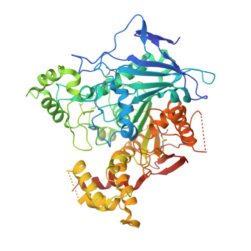
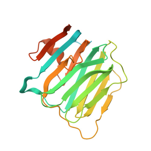
Crystal structure of the Neuroligin-1/Neurexin-1beta synaptic adhesion complex


Experimental Data Snapshot

  • Method: X-RAY DIFFRACTION
  • Resolution: 3.50 Å
  • R-Value Free: 
    0.276 (Depositor), 0.277 (DCC) 
  • R-Value Work: 
    0.246 (Depositor), 0.248 (DCC) 

wwPDB Validation   3D Report Full Report


Ligand Structure Quality Assessment 

Created with Raphaël 2.3.0Worse 01 BetterLigand structure goodness of fit to experimental dataBest fitted NAGClick on this verticalbar to view details

This is version 2.1 of the entry. See complete history


Literature

Structures of Neuroligin-1 and the Neuroligin-1/Neurexin-1beta Complex Reveal Specific Protein-Protein and Protein-Ca(2+) Interactions.

Arac, D.Boucard, A.A.Ozkan, E.Strop, P.Newell, E.Sudhof, T.C.Brunger, A.T.

(2007) Neuron 56: 992-1003

  • DOI: https://doi.org/10.1016/j.neuron.2007.12.002
  • Primary Citation of Related Structures:  
    3BIW, 3BIX

  • PubMed Abstract: 

    Neurexins and neuroligins provide trans-synaptic connectivity by the Ca2+-dependent interaction of their alternatively spliced extracellular domains. Neuroligins specify synapses in an activity-dependent manner, presumably by binding to neurexins. Here, we present the crystal structures of neuroligin-1 in isolation and in complex with neurexin-1 beta. Neuroligin-1 forms a constitutive dimer, and two neurexin-1 beta monomers bind to two identical surfaces on the opposite faces of the neuroligin-1 dimer to form a heterotetramer. The neuroligin-1/neurexin-1 beta complex exhibits a nanomolar affinity and includes a large binding interface that contains bound Ca2+. Alternatively spliced sites in neurexin-1 beta and in neuroligin-1 are positioned nearby the binding interface, explaining how they regulate the interaction. Structure-based mutations of neuroligin-1 at the interface disrupt binding to neurexin-1 beta, but not the folding of neuroligin-1 and confirm the validity of the binding interface of the neuroligin-1/neurexin-1 beta complex. Our results provide molecular insights for understanding the role of cell-adhesion proteins in synapse function.


  • Organizational Affiliation

    Howard Hughes Medical Institute, Stanford University, Stanford, CA 94305, USA.


Macromolecules
Find similar proteins by:  (by identity cutoff)  |  3D Structure
Entity ID: 1
MoleculeChains Sequence LengthOrganismDetailsImage
Neuroligin-1
A, B, C, D
574Rattus norvegicusMutation(s): 0 
Gene Names: Nlgn1
Membrane Entity: Yes 
UniProt
Find proteins for Q62765 (Rattus norvegicus)
Explore Q62765 
Go to UniProtKB:  Q62765
Entity Groups  
Sequence Clusters30% Identity50% Identity70% Identity90% Identity95% Identity100% Identity
UniProt GroupQ62765
Glycosylation
Glycosylation Sites: 3Go to GlyGen: Q62765-1
Sequence Annotations
Expand
  • Reference Sequence
Find similar proteins by:  (by identity cutoff)  |  3D Structure
Entity ID: 2
MoleculeChains Sequence LengthOrganismDetailsImage
Neurexin-1-beta
E, F, G, H
243Rattus norvegicusMutation(s): 0 
Gene Names: Nrxn1
Membrane Entity: Yes 
UniProt
Find proteins for Q63372 (Rattus norvegicus)
Explore Q63372 
Go to UniProtKB:  Q63372
Entity Groups  
Sequence Clusters30% Identity50% Identity70% Identity90% Identity95% Identity100% Identity
UniProt GroupQ63372
Glycosylation
Glycosylation Sites: 1Go to GlyGen: Q63372-2
Sequence Annotations
Expand
  • Reference Sequence
Oligosaccharides

Help

Entity ID: 3
MoleculeChains Length2D Diagram Glycosylation3D Interactions
2-acetamido-2-deoxy-beta-D-glucopyranose-(1-4)-2-acetamido-2-deoxy-beta-D-glucopyranose
I, J, K
2N-Glycosylation
Glycosylation Resources
GlyTouCan:  G42666HT
GlyCosmos:  G42666HT
GlyGen:  G42666HT
Small Molecules
Ligands 2 Unique
IDChains Name / Formula / InChI Key2D Diagram3D Interactions
NAG
Query on NAG

Download Ideal Coordinates CCD File 
L [auth A]
M [auth A]
N [auth A]
O [auth B]
P [auth B]
2-acetamido-2-deoxy-beta-D-glucopyranose
C8 H15 N O6
OVRNDRQMDRJTHS-FMDGEEDCSA-N
CA
Query on CA

Download Ideal Coordinates CCD File 
AA [auth F]
BA [auth G]
CA [auth G]
X [auth E]
Y [auth E]
CALCIUM ION
Ca
BHPQYMZQTOCNFJ-UHFFFAOYSA-N
Experimental Data & Validation

Experimental Data

  • Method: X-RAY DIFFRACTION
  • Resolution: 3.50 Å
  • R-Value Free:  0.276 (Depositor), 0.277 (DCC) 
  • R-Value Work:  0.246 (Depositor), 0.248 (DCC) 
Space Group: C 1 2 1
Unit Cell:
Length ( Å )Angle ( ˚ )
a = 229.83α = 90
b = 148.796β = 90.38
c = 123.602γ = 90
Software Package:
Software NamePurpose
DENZOdata reduction
SCALEPACKdata scaling
PHASERphasing
CNSrefinement
PDB_EXTRACTdata extraction
HKL-2000data reduction
HKL-2000data scaling

Structure Validation

View Full Validation Report


Ligand Structure Quality Assessment 

Created with Raphaël 2.3.0Worse 01 BetterLigand structure goodness of fit to experimental dataBest fitted NAGClick on this verticalbar to view details

Entry History 

Deposition Data

Revision History  (Full details and data files)

  • Version 1.0: 2007-12-18
    Type: Initial release
  • Version 1.1: 2011-07-13
    Changes: Non-polymer description, Version format compliance
  • Version 1.2: 2017-08-23
    Changes: Refinement description, Source and taxonomy
  • Version 1.3: 2017-10-25
    Changes: Refinement description
  • Version 2.0: 2020-07-29
    Type: Remediation
    Reason: Carbohydrate remediation
    Changes: Atomic model, Data collection, Database references, Derived calculations, Structure summary
  • Version 2.1: 2024-10-16
    Changes: Data collection, Database references, Structure summary