3C0N

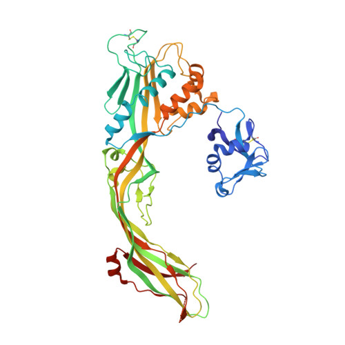
Crystal structure of the proaerolysin mutant Y221G at 2.2 A


Experimental Data Snapshot

  • Method: X-RAY DIFFRACTION
  • Resolution: 2.20 Å
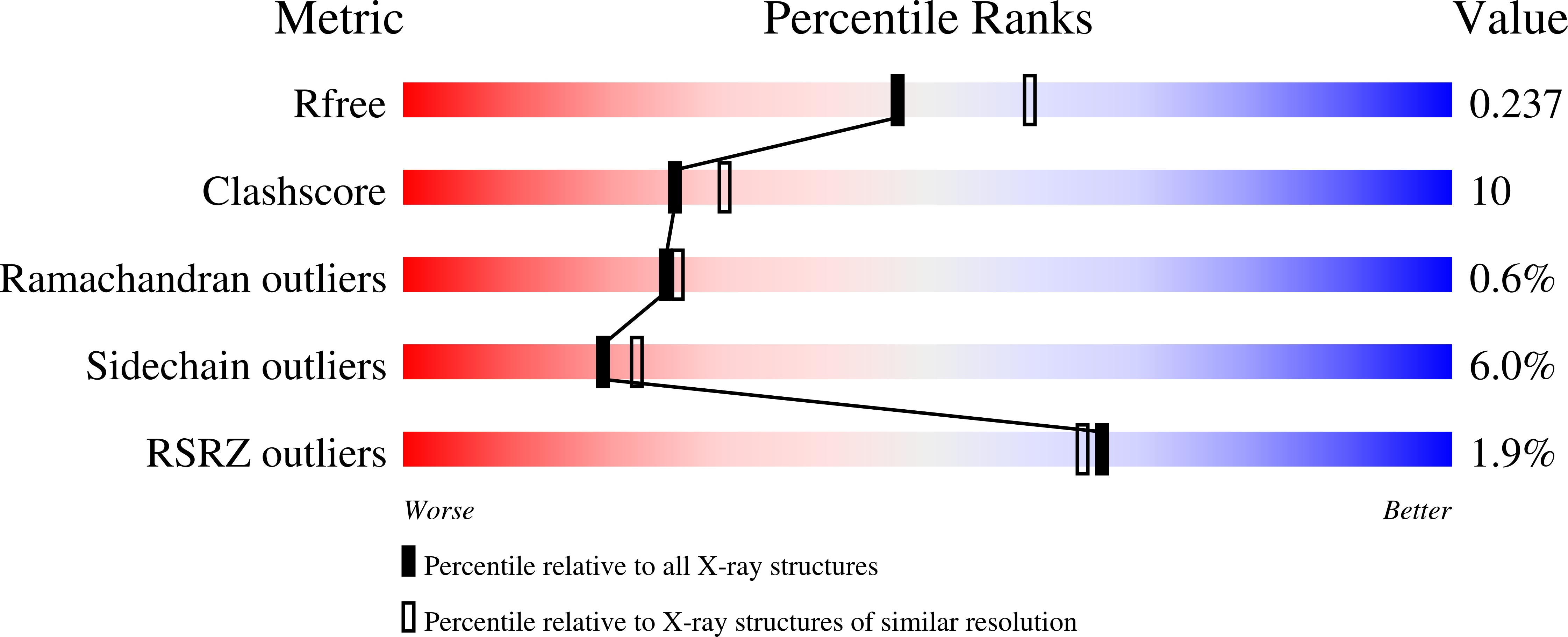
  • R-Value Free: 0.238 
  • R-Value Work: 0.189 
  • R-Value Observed: 0.192 

Starting Model: experimental
View more details

wwPDB Validation   3D Report Full Report


This is version 1.5 of the entry. See complete history


Literature

Molecular assembly of the aerolysin pore reveals a swirling membrane-insertion mechanism.

Degiacomi, M.T.Iacovache, I.Pernot, L.Chami, M.Kudryashev, M.Stahlberg, H.van der Goot, F.G.Dal Peraro, M.

(2013) Nat Chem Biol 9: 623-629

  • DOI: https://doi.org/10.1038/nchembio.1312
  • Primary Citation of Related Structures:  
    3C0M, 3C0N, 3C0O

  • PubMed Abstract: 

    Aerolysin is the founding member of a superfamily of β-pore-forming toxins whose pore structure is unknown. We have combined X-ray crystallography, cryo-EM, molecular dynamics and computational modeling to determine the structures of aerolysin mutants in their monomeric and heptameric forms, trapped at various stages of the pore formation process. A dynamic modeling approach based on swarm intelligence was applied, whereby the intrinsic flexibility of aerolysin extracted from new X-ray structures was used to fully exploit the cryo-EM spatial restraints. Using this integrated strategy, we obtained a radically new arrangement of the prepore conformation and a near-atomistic structure of the aerolysin pore, which is fully consistent with all of the biochemical data available so far. Upon transition from the prepore to pore, the aerolysin heptamer shows a unique concerted swirling movement, accompanied by a vertical collapse of the complex, ultimately leading to the insertion of a transmembrane β-barrel.


  • Organizational Affiliation

    1] Institute of Bioengineering, School of Life Sciences, Ecole Polytechnique Fédérale de Lausanne, Lausanne, Switzerland. [2] [3].


Macromolecules
Find similar proteins by:  (by identity cutoff)  |  3D Structure
Entity ID: 1
MoleculeChains Sequence LengthOrganismDetailsImage
Aerolysin
A, B
470Aeromonas hydrophilaMutation(s): 1 
Gene Names: aerA
Membrane Entity: Yes 
UniProt
Find proteins for P09167 (Aeromonas hydrophila)
Explore P09167 
Go to UniProtKB:  P09167
Entity Groups  
Sequence Clusters30% Identity50% Identity70% Identity90% Identity95% Identity100% Identity
UniProt GroupP09167
Sequence Annotations
Expand
  • Reference Sequence
Experimental Data & Validation

Experimental Data

  • Method: X-RAY DIFFRACTION
  • Resolution: 2.20 Å
  • R-Value Free: 0.238 
  • R-Value Work: 0.189 
  • R-Value Observed: 0.192 
  • Space Group: P 21 21 21
Unit Cell:
Length ( Å )Angle ( ˚ )
a = 70.36α = 90
b = 89.768β = 90
c = 166.616γ = 90
Software Package:
Software NamePurpose
REFMACrefinement
MOSFLMdata reduction
SCALAdata scaling
PHASERphasing

Structure Validation

View Full Validation Report


Entry History 

Deposition Data

Revision History  (Full details and data files)

  • Version 1.0: 2008-02-12
    Type: Initial release
  • Version 1.1: 2011-07-13
    Changes: Version format compliance
  • Version 1.2: 2013-10-02
    Changes: Database references
  • Version 1.3: 2021-11-10
    Changes: Database references
  • Version 1.4: 2023-11-01
    Changes: Data collection, Refinement description
  • Version 1.5: 2024-10-30
    Changes: Structure summary