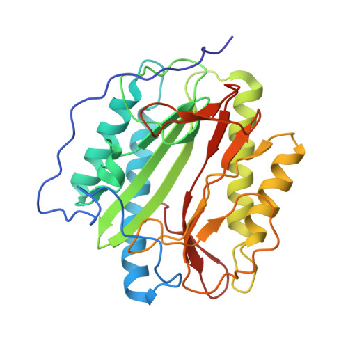
3PKA

M. tuberculosis MetAP with bengamide analog Y02, in Mn form


Experimental Data Snapshot

  • Method: X-RAY DIFFRACTION
  • Resolution: 1.25 Å
  • R-Value Free: 0.188 
  • R-Value Work: 0.169 
  • R-Value Observed: 0.169 

wwPDB Validation   3D Report Full Report


Ligand Structure Quality Assessment 


This is version 1.3 of the entry. See complete history


Literature

Inhibition of Mycobacterium tuberculosis Methionine Aminopeptidases by Bengamide Derivatives.

Lu, J.P.Yuan, X.H.Yuan, H.Wang, W.L.Wan, B.Franzblau, S.G.Ye, Q.Z.

(2011) ChemMedChem 6: 1041-1048

  • DOI: https://doi.org/10.1002/cmdc.201100003
  • Primary Citation of Related Structures:  
    3PKA, 3PKB, 3PKC, 3PKD, 3PKE

  • PubMed Abstract: 

    Methionine aminopeptidase (MetAP) carries out an essential function of protein N-terminal processing in many bacteria and is a promising target for the development of novel antitubercular agents. Natural bengamides potently inhibit the proliferation of mammalian cells by targeting MetAP enzymes, and the X-ray crystal structure of human type 2 MetAP in complex with a bengamide derivative reveals the key interactions at the active site. By preserving the interactions with the conserved residues inside the binding pocket while exploring the differences between bacterial and human MetAPs around the binding pocket, seven bengamide derivatives were synthesized and evaluated for inhibition of MtMetAP1a and MtMetAP1c in different metalloforms, inhibition of M. tuberculosis growth in replicating and non-replicating states, and inhibition of human K562 cell growth. Potent inhibition of MtMetAP1a and MtMetAP1c and modest growth inhibition of M. tuberculosis were observed for some of these derivatives. Crystal structures of MtMetAP1c in complex with two of the derivatives provided valuable structural information for improvement of these inhibitors for potency and selectivity.


  • Organizational Affiliation

    Department of Biochemistry and Molecular Biology, Indiana University School of Medicine, 635 Barnhill Drive, Indianapolis, IN 46202, USA.


Macromolecules
Find similar proteins by:  (by identity cutoff)  |  3D Structure
Entity ID: 1
MoleculeChains Sequence LengthOrganismDetailsImage
Methionine aminopeptidase285Mycobacterium tuberculosisMutation(s): 0 
Gene Names: mapmapBMT2929MTV003.07cRv2861c
EC: 3.4.11.18
UniProt
Find proteins for P9WK19 (Mycobacterium tuberculosis (strain ATCC 25618 / H37Rv))
Explore P9WK19 
Go to UniProtKB:  P9WK19
Entity Groups  
Sequence Clusters30% Identity50% Identity70% Identity90% Identity95% Identity100% Identity
UniProt GroupP9WK19
Sequence Annotations
Expand
  • Reference Sequence
Small Molecules
Ligands 4 Unique
IDChains Name / Formula / InChI Key2D Diagram3D Interactions
Y02
Query on Y02

Download Ideal Coordinates CCD File 
D [auth A](2R,3R,4S,5R,6E)-3,4,5-trihydroxy-2-methoxy-8,8-dimethyl-N-[2-(2,4,6-trimethylphenoxy)ethyl]non-6-enamide
C23 H37 N O6
UMRMEUOEFMNZRM-SWQIVCBNSA-N
SO4
Query on SO4

Download Ideal Coordinates CCD File 
E [auth A]SULFATE ION
O4 S
QAOWNCQODCNURD-UHFFFAOYSA-L
MN
Query on MN

Download Ideal Coordinates CCD File 
B [auth A],
C [auth A]
MANGANESE (II) ION
Mn
WAEMQWOKJMHJLA-UHFFFAOYSA-N
CL
Query on CL

Download Ideal Coordinates CCD File 
F [auth A]CHLORIDE ION
Cl
VEXZGXHMUGYJMC-UHFFFAOYSA-M
Binding Affinity Annotations 
IDSourceBinding Affinity
Y02 PDBBind:  3PKA IC50: 540 (nM) from 1 assay(s)
Experimental Data & Validation

Experimental Data

  • Method: X-RAY DIFFRACTION
  • Resolution: 1.25 Å
  • R-Value Free: 0.188 
  • R-Value Work: 0.169 
  • R-Value Observed: 0.169 
  • Space Group: P 63
Unit Cell:
Length ( Å )Angle ( ˚ )
a = 106.003α = 90
b = 106.003β = 90
c = 50.395γ = 120
Software Package:
Software NamePurpose
MOLREPphasing
REFMACrefinement
PDB_EXTRACTdata extraction
HKL-3000data collection
HKL-3000data reduction
HKL-3000data scaling

Structure Validation

View Full Validation Report


Ligand Structure Quality Assessment 


Entry History 

Deposition Data

Revision History  (Full details and data files)

  • Version 1.0: 2011-04-20
    Type: Initial release
  • Version 1.1: 2011-07-13
    Changes: Version format compliance
  • Version 1.2: 2013-05-29
    Changes: Non-polymer description
  • Version 1.3: 2024-02-21
    Changes: Data collection, Database references, Derived calculations