


Crystal Structure of PF10_0086, adenylate kinase from plasmodium falciparum
Wernimont, A.K., Loppnau, P., Crombet, L., Weadge, J., Perieteanu, A., Edwards, A.M., Arrowsmith, C.H., Park, H., Bountra, C., Hui, R., Amani, M.To be published.
Experimental Data Snapshot
Entity ID: 1 | |||||
|---|---|---|---|---|---|
| Molecule | Chains | Sequence Length | Organism | Details | Image |
| Adenylate kinase 2 | 243 | Plasmodium falciparum | Mutation(s): 0 EC: 2.7.4.3 | 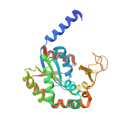 | |
UniProt | |||||
Find proteins for Q8IJV6 (Plasmodium falciparum (isolate 3D7)) Explore Q8IJV6 Go to UniProtKB: Q8IJV6 | |||||
Entity Groups | |||||
| Sequence Clusters | 30% Identity50% Identity70% Identity90% Identity95% Identity100% Identity | ||||
| UniProt Group | Q8IJV6 | ||||
Sequence AnnotationsExpand | |||||
| |||||
| Ligands 4 Unique | |||||
|---|---|---|---|---|---|
| ID | Chains | Name / Formula / InChI Key | 2D Diagram | 3D Interactions | |
| ATP Query on ATP | K [auth D] | ADENOSINE-5'-TRIPHOSPHATE C10 H16 N5 O13 P3 ZKHQWZAMYRWXGA-KQYNXXCUSA-N |  | ||
| ADP Query on ADP | H [auth A], I [auth B], J [auth B] | ADENOSINE-5'-DIPHOSPHATE C10 H15 N5 O10 P2 XTWYTFMLZFPYCI-KQYNXXCUSA-N |  | ||
| AMP Query on AMP | G [auth A] | ADENOSINE MONOPHOSPHATE C10 H14 N5 O7 P UDMBCSSLTHHNCD-KQYNXXCUSA-N |  | ||
| MG Query on MG | E [auth A], F [auth A] | MAGNESIUM ION Mg JLVVSXFLKOJNIY-UHFFFAOYSA-N |  | ||
| Length ( Å ) | Angle ( ˚ ) |
|---|---|
| a = 81.395 | α = 90 |
| b = 76.663 | β = 100.78 |
| c = 93.768 | γ = 90 |
| Software Name | Purpose |
|---|---|
| DENZO | data reduction |
| SCALEPACK | data scaling |
| PHASER | phasing |
| BUSTER-TNT | refinement |
| PDB_EXTRACT | data extraction |
| HKL-3000 | data collection |
| BUSTER | refinement |


