3V6O | pdb_00003v6o

Leptin Receptor-antibody complex


Experimental Data Snapshot

  • Method: X-RAY DIFFRACTION
  • Resolution: 1.95 Å
  • R-Value Free: 
    0.213 (Depositor), 0.220 (DCC) 
  • R-Value Work: 
    0.171 (Depositor), 0.180 (DCC) 
  • R-Value Observed: 
    0.173 (Depositor) 

wwPDB Validation   3D Report Full Report


Ligand Structure Quality Assessment 

Created with Raphaël 2.3.0Worse 01 BetterLigand structure goodness of fit to experimental dataBest fitted NAGClick on this verticalbar to view details

This is version 1.4 of the entry. See complete history


Literature

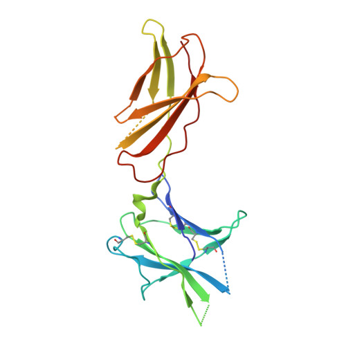
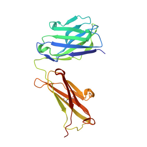
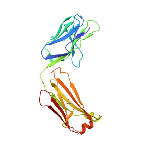
Structure of the human obesity receptor leptin-binding domain reveals the mechanism of leptin antagonism by a monoclonal antibody.

Carpenter, B.Hemsworth, G.R.Wu, Z.Maamra, M.Strasburger, C.J.Ross, R.J.Artymiuk, P.J.

(2012) Structure 20: 487-497

  • DOI: https://doi.org/10.1016/j.str.2012.01.019
  • Primary Citation of Related Structures:  
    3V6O, 3VG0

  • PubMed Abstract: 

    Leptin regulates energy homeostasis, fertility, and the immune system, making it an important drug target. However, due to a complete lack of structural data for the obesity receptor (ObR), leptin's mechanism of receptor activation remains poorly understood. We have crystallized the Fab fragment of a leptin-blocking monoclonal antibody (9F8), both in its uncomplexed state and bound to the leptin-binding domain (LBD) of human ObR. We describe the structure of the LBD-9F8 Fab complex and the conformational changes in 9F8 associated with LBD binding. A molecular model of the putative leptin-LBD complex reveals that 9F8 Fab blocks leptin binding through only a small (10%) overlap in their binding sites, and that leptin binding is likely to involve an induced fit mechanism. This crystal structure of the leptin-binding domain of the obesity receptor will facilitate the design of therapeutics to modulate leptin signaling.


  • Organizational Affiliation

    Academic Unit of Diabetes, Endocrinology and Reproduction, Department of Human Metabolism, University of Sheffield, Sheffield S10 2JF, UK.


Macromolecules
Find similar proteins by:  (by identity cutoff)  |  3D Structure
Entity ID: 1
MoleculeChains Sequence LengthOrganismDetailsImage
Leptin receptor
A, B
206Homo sapiensMutation(s): 0 
Gene Names: DBLEPROBR
UniProt & NIH Common Fund Data Resources
Find proteins for P48357 (Homo sapiens)
Explore P48357 
Go to UniProtKB:  P48357
PHAROS:  P48357
GTEx:  ENSG00000116678 
Entity Groups  
Sequence Clusters30% Identity50% Identity70% Identity90% Identity95% Identity100% Identity
UniProt GroupP48357
Sequence Annotations
Expand
  • Reference Sequence
Find similar proteins by:  (by identity cutoff)  |  3D Structure
Entity ID: 2
MoleculeChains Sequence LengthOrganismDetailsImage
Monoclonal antibody 9F8 fab fragment Heavy chain
C, D
221Mus musculusMutation(s): 0 
Entity Groups  
Sequence Clusters30% Identity50% Identity70% Identity90% Identity95% Identity100% Identity
Sequence Annotations
Expand
  • Reference Sequence
Find similar proteins by:  (by identity cutoff)  |  3D Structure
Entity ID: 3
MoleculeChains Sequence LengthOrganismDetailsImage
Monoclonal antibody 9F8 fab fragment Light chain
E, F
215Mus musculusMutation(s): 0 
Entity Groups  
Sequence Clusters30% Identity50% Identity70% Identity90% Identity95% Identity100% Identity
Glycosylation
Glycosylation Sites: 1
Sequence Annotations
Expand
  • Reference Sequence
Small Molecules
Ligands 5 Unique
IDChains Name / Formula / InChI Key2D Diagram3D Interactions
NAG
Query on NAG

Download Ideal Coordinates CCD File 
MA [auth F]2-acetamido-2-deoxy-beta-D-glucopyranose
C8 H15 N O6
OVRNDRQMDRJTHS-FMDGEEDCSA-N
CYS
Query on CYS

Download Ideal Coordinates CCD File 
G [auth A],
P [auth B]
CYSTEINE
C3 H7 N O2 S
XUJNEKJLAYXESH-REOHCLBHSA-N
EDO
Query on EDO

Download Ideal Coordinates CCD File 
BA [auth D]
CA [auth D]
DA [auth D]
EA [auth D]
FA [auth D]
1,2-ETHANEDIOL
C2 H6 O2
LYCAIKOWRPUZTN-UHFFFAOYSA-N
ACT
Query on ACT

Download Ideal Coordinates CCD File 
AA [auth D]
IA [auth E]
NA [auth F]
OA [auth F]
V [auth C]
ACETATE ION
C2 H3 O2
QTBSBXVTEAMEQO-UHFFFAOYSA-M
NA
Query on NA

Download Ideal Coordinates CCD File 
Z [auth C]SODIUM ION
Na
FKNQFGJONOIPTF-UHFFFAOYSA-N
Experimental Data & Validation

Experimental Data

  • Method: X-RAY DIFFRACTION
  • Resolution: 1.95 Å
  • R-Value Free:  0.213 (Depositor), 0.220 (DCC) 
  • R-Value Work:  0.171 (Depositor), 0.180 (DCC) 
  • R-Value Observed: 0.173 (Depositor) 
Space Group: P 21 21 21
Unit Cell:
Length ( Å )Angle ( ˚ )
a = 89.814α = 90
b = 118.829β = 90
c = 171.281γ = 90
Software Package:
Software NamePurpose
REFMACrefinement
PDB_EXTRACTdata extraction
ADSCdata collection
MOSFLMdata reduction
SCALAdata scaling
PHASERphasing

Structure Validation

View Full Validation Report


Ligand Structure Quality Assessment 

Created with Raphaël 2.3.0Worse 01 BetterLigand structure goodness of fit to experimental dataBest fitted NAGClick on this verticalbar to view details

Entry History 

Deposition Data

Revision History  (Full details and data files)

  • Version 1.0: 2012-03-14
    Type: Initial release
  • Version 1.1: 2012-04-25
    Changes: Database references
  • Version 1.2: 2020-07-29
    Type: Remediation
    Reason: Carbohydrate remediation
    Changes: Data collection, Database references, Derived calculations, Structure summary
  • Version 1.3: 2023-12-27
    Changes: Data collection, Database references, Derived calculations, Structure summary
  • Version 1.4: 2024-10-16
    Changes: Structure summary