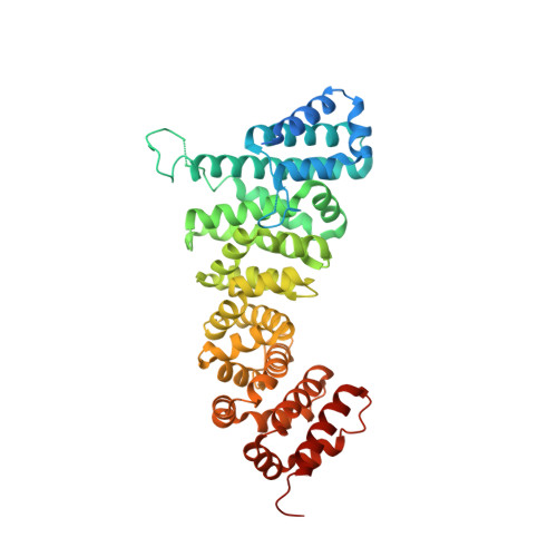
3NMZ

Crystal structure of APC complexed with Asef


Experimental Data Snapshot

  • Method: X-RAY DIFFRACTION
  • Resolution: 3.01 Å
  • R-Value Free: 0.284 
  • R-Value Work: 0.249 
  • R-Value Observed: 0.251 

Starting Models: experimental
View more details

wwPDB Validation   3D Report Full Report


This is version 1.5 of the entry. See complete history


Literature

Structural basis for the recognition of Asef by adenomatous polyposis coli.

Zhang, Z.Chen, L.Gao, L.Lin, K.Zhu, L.Lu, Y.Shi, X.Gao, Y.Zhou, J.Xu, P.Zhang, J.Wu, G.

(2012) Cell Res 22: 372-386

  • DOI: https://doi.org/10.1038/cr.2011.119
  • Primary Citation of Related Structures:  
    3NMW, 3NMX, 3NMZ

  • PubMed Abstract: 

    Adenomatous polyposis coli (APC) regulates cell-cell adhesion and cell migration through activating the APC-stimulated guanine nucleotide-exchange factor (GEF; Asef), which is usually autoinhibited through the binding between its Src homology 3 (SH3) and Dbl homology (DH) domains. The APC-activated Asef stimulates the small GTPase Cdc42, which leads to decreased cell-cell adherence and enhanced cell migration. In colorectal cancers, truncated APC constitutively activates Asef and promotes cancer cell migration and angiogenesis. Here, we report crystal structures of the human APC/Asef complex. We find that the armadillo repeat domain of APC uses a highly conserved surface groove to recognize the APC-binding region (ABR) of Asef, conformation of which changes dramatically upon binding to APC. Key residues on APC and Asef for the complex formation were mutated and their importance was demonstrated by binding and activity assays. Structural superimposition of the APC/Asef complex with autoinhibited Asef suggests that the binding between APC and Asef might create a steric clash between Asef-DH domain and APC, which possibly leads to a conformational change in Asef that stimulates its GEF activity. Our structures thus elucidate the molecular mechanism of Asef recognition by APC, as well as provide a potential target for pharmaceutical intervention against cancers.


  • Organizational Affiliation

    State Key Laboratory of Microbial Metabolism, Shanghai Jiao Tong University, Shanghai 200240, China.


Macromolecules
Find similar proteins by:  (by identity cutoff)  |  3D Structure
Entity ID: 1
MoleculeChains Sequence LengthOrganismDetailsImage
APC variant protein
A, B
458Homo sapiensMutation(s): 0 
Gene Names: APC variant protein
UniProt & NIH Common Fund Data Resources
Find proteins for P25054 (Homo sapiens)
Explore P25054 
Go to UniProtKB:  P25054
PHAROS:  P25054
GTEx:  ENSG00000134982 
Entity Groups  
Sequence Clusters30% Identity50% Identity70% Identity90% Identity95% Identity100% Identity
UniProt GroupP25054
Sequence Annotations
Expand
  • Reference Sequence
Find similar proteins by:  (by identity cutoff)  |  3D Structure
Entity ID: 2
MoleculeChains Sequence LengthOrganismDetailsImage
Rho guanine nucleotide exchange factor 4C [auth D],
D [auth C]
116Homo sapiensMutation(s): 0 
Gene Names: ARHGEF4KIAA1112
UniProt & NIH Common Fund Data Resources
Find proteins for Q9NR80 (Homo sapiens)
Explore Q9NR80 
Go to UniProtKB:  Q9NR80
PHAROS:  Q9NR80
GTEx:  ENSG00000136002 
Entity Groups  
Sequence Clusters30% Identity50% Identity70% Identity90% Identity95% Identity100% Identity
UniProt GroupQ9NR80
Sequence Annotations
Expand
  • Reference Sequence
Experimental Data & Validation

Experimental Data

  • Method: X-RAY DIFFRACTION
  • Resolution: 3.01 Å
  • R-Value Free: 0.284 
  • R-Value Work: 0.249 
  • R-Value Observed: 0.251 
  • Space Group: H 3
Unit Cell:
Length ( Å )Angle ( ˚ )
a = 163.175α = 90
b = 163.175β = 90
c = 242.593γ = 120
Software Package:
Software NamePurpose
SCALEPACKdata scaling
PHASERphasing
REFMACrefinement
PDB_EXTRACTdata extraction

Structure Validation

View Full Validation Report


Entry History 

Deposition Data

Revision History  (Full details and data files)

  • Version 1.0: 2011-07-06
    Type: Initial release
  • Version 1.1: 2011-07-13
    Changes: Version format compliance
  • Version 1.2: 2016-12-21
    Changes: Structure summary
  • Version 1.3: 2017-03-15
    Changes: Database references
  • Version 1.4: 2019-12-25
    Changes: Database references
  • Version 1.5: 2023-11-01
    Changes: Data collection, Database references, Refinement description