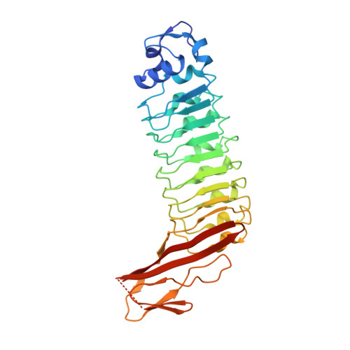
4AW4

Engineered variant of Listeria monocytogenes InlB internalin domain with an additional leucine rich repeat inserted


Experimental Data Snapshot

  • Method: X-RAY DIFFRACTION
  • Resolution: 1.93 Å
  • R-Value Free: 0.205 
  • R-Value Work: 0.167 
  • R-Value Observed: 0.168 

Starting Model: experimental
View more details

wwPDB Validation   3D Report Full Report


This is version 1.6 of the entry. See complete history


Literature

Engineered Variants of Inlb with an Additional Leucine-Rich Repeat Discriminate between Physiologically Relevant and Packing Contacts in Crystal Structures of the Inlb:Met Complex.

Niemann, H.H.Gherardi, E.Bleymuller, W.M.Heinz, D.W.

(2012) Protein Sci 21: 1528

  • DOI: https://doi.org/10.1002/pro.2142
  • Primary Citation of Related Structures:  
    4AW4

  • PubMed Abstract: 

    The physiological relevance of contacts in crystal lattices often remains elusive. This was also the case for the complex between the invasion protein internalin B (InlB) from Listeria monocytogenes and its host cell receptor, the human receptor tyrosine kinase (RTK) MET. InlB is a MET agonist and induces bacterial host cell invasion. Activation of RTKs generally involves ligand-induced dimerization of the receptor ectodomain. The two currently available crystal structures of the InlB:MET complex show the same arrangement of InlB and MET in a 1:1 complex, but different dimeric 2:2 assemblies. Only one of these 2:2 assemblies is predicted to be stable by a computational procedure. This assembly is mainly stabilized by a contact between the Cap domain of InlB from one and the Sema domain of MET from another 1:1 complex. Here, we probe the physiological relevance of this interaction. We generated variants of the leucine-rich repeat (LRR) protein InlB by inserting an additional repeat between the first and the second LRR. This should allow formation of the 1:1 complex but disrupt the potential 2:2 complex involving the Cap-Sema contact due to steric distortions. A crystal structure of one of the engineered proteins showed that it folded properly. Binding affinity to MET was comparable to that of wild-type InlB. The InlB variant induced MET phosphorylation and cell scatter like wild-type InlB. These results suggest that the Cap-Sema interaction is not physiologically relevant and support the previously proposed assembly, in which a 2:2 InlB:MET complex is built around a ligand dimer.


  • Organizational Affiliation

    Department of Chemistry, Bielefeld University, 33501 Bielefeld, Germany. Hartmut.Niemann@uni-bielefeld.de


Macromolecules
Find similar proteins by:  (by identity cutoff)  |  3D Structure
Entity ID: 1
MoleculeChains Sequence LengthOrganismDetailsImage
INTERNALIN B
A, B, C
311Listeria monocytogenes EGD-eMutation(s): 0 
UniProt
Find proteins for P0DQD3 (Listeria monocytogenes serotype 1/2a (strain EGD / Mackaness))
Explore P0DQD3 
Go to UniProtKB:  P0DQD3
Entity Groups  
Sequence Clusters30% Identity50% Identity70% Identity90% Identity95% Identity100% Identity
UniProt GroupP0DQD3
Sequence Annotations
Expand
  • Reference Sequence
Small Molecules
Ligands 2 Unique
IDChains Name / Formula / InChI Key2D Diagram3D Interactions
SO4
Query on SO4

Download Ideal Coordinates CCD File 
AA [auth B]
BA [auth B]
CA [auth C]
D [auth A]
E [auth A]
AA [auth B],
BA [auth B],
CA [auth C],
D [auth A],
E [auth A],
F [auth A],
FA [auth C],
G [auth A],
GA [auth C],
H [auth A],
HA [auth C],
I [auth A],
IA [auth C],
JA [auth C],
O [auth A],
P [auth A],
Q [auth A],
S [auth B],
T [auth B],
X [auth B],
Y [auth B],
Z [auth B]
SULFATE ION
O4 S
QAOWNCQODCNURD-UHFFFAOYSA-L
GOL
Query on GOL

Download Ideal Coordinates CCD File 
DA [auth C]
EA [auth C]
J [auth A]
K [auth A]
L [auth A]
DA [auth C],
EA [auth C],
J [auth A],
K [auth A],
L [auth A],
M [auth A],
N [auth A],
R [auth B],
U [auth B],
V [auth B],
W [auth B]
GLYCEROL
C3 H8 O3
PEDCQBHIVMGVHV-UHFFFAOYSA-N
Experimental Data & Validation

Experimental Data

  • Method: X-RAY DIFFRACTION
  • Resolution: 1.93 Å
  • R-Value Free: 0.205 
  • R-Value Work: 0.167 
  • R-Value Observed: 0.168 
  • Space Group: C 1 2 1
Unit Cell:
Length ( Å )Angle ( ˚ )
a = 179.08α = 90
b = 102.15β = 90.23
c = 69.81γ = 90
Software Package:
Software NamePurpose
REFMACrefinement
XDSdata reduction
XSCALEdata scaling
PHASERphasing

Structure Validation

View Full Validation Report


Entry History 

Deposition Data

Revision History  (Full details and data files)

  • Version 1.0: 2012-08-15
    Type: Initial release
  • Version 1.1: 2012-08-29
    Changes: Database references
  • Version 1.2: 2012-10-03
    Changes: Database references
  • Version 1.3: 2019-05-08
    Changes: Data collection, Derived calculations, Experimental preparation, Other
  • Version 1.4: 2019-05-22
    Changes: Data collection, Experimental preparation
  • Version 1.5: 2019-07-17
    Changes: Data collection
  • Version 1.6: 2023-12-20
    Changes: Data collection, Database references, Derived calculations, Other, Refinement description