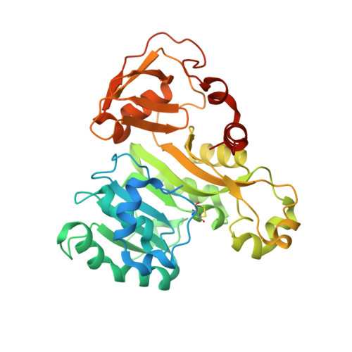
4FR0

ArsM arsenic(III) S-adenosylmethionine methyltransferase with SAM


Experimental Data Snapshot

  • Method: X-RAY DIFFRACTION
  • Resolution: 2.75 Å
  • R-Value Free: 0.283 
  • R-Value Work: 0.206 
  • R-Value Observed: 0.209 

Starting Model: experimental
View more details

wwPDB Validation   3D Report Full Report


Ligand Structure Quality Assessment 


This is version 1.3 of the entry. See complete history


Literature

Structure of an As(III) S-Adenosylmethionine Methyltransferase: Insights into the Mechanism of Arsenic Biotransformation.

Ajees, A.A.Marapakala, K.Packianathan, C.Sankaran, B.Rosen, B.P.

(2012) Biochemistry 51: 5476-5485

  • DOI: https://doi.org/10.1021/bi3004632
  • Primary Citation of Related Structures:  
    4FR0, 4FS8, 4FSD

  • PubMed Abstract: 

    Enzymatic methylation of arsenic is a detoxification process in microorganisms but in humans may activate the metalloid to more carcinogenic species. We describe the first structure of an As(III) S-adenosylmethionine methyltransferase by X-ray crystallography that reveals a novel As(III) binding domain. The structure of the methyltransferase from the thermophilic eukaryotic alga Cyanidioschyzon merolae reveals the relationship between the arsenic and S-adenosylmethionine binding sites to a final resolution of ∼1.6 Å. As(III) binding causes little change in conformation, but binding of SAM reorients helix α4 and a loop (residues 49-80) toward the As(III) binding domain, positioning the methyl group for transfer to the metalloid. There is no evidence of a reductase domain. These results are consistent with previous suggestions that arsenic remains trivalent during the catalytic cycle. A homology model of human As(III) S-adenosylmethionine methyltransferase with the location of known polymorphisms was constructed. The structure provides insights into the mechanism of substrate binding and catalysis.


  • Organizational Affiliation

    Department of Cellular Biology and Pharmacology, Herbert Wertheim College of Medicine, Florida International University, Miami, 33199, United States.


Macromolecules
Find similar proteins by:  (by identity cutoff)  |  3D Structure
Entity ID: 1
MoleculeChains Sequence LengthOrganismDetailsImage
Arsenic methyltransferase383Cyanidioschyzon sp. 5508Mutation(s): 0 
Gene Names: arsM
UniProt
Find proteins for C0JV69 (Cyanidioschyzon sp. 5508)
Explore C0JV69 
Go to UniProtKB:  C0JV69
Entity Groups  
Sequence Clusters30% Identity50% Identity70% Identity90% Identity95% Identity100% Identity
UniProt GroupC0JV69
Sequence Annotations
Expand
  • Reference Sequence
Small Molecules
Ligands 1 Unique
IDChains Name / Formula / InChI Key2D Diagram3D Interactions
SAM
Query on SAM

Download Ideal Coordinates CCD File 
B [auth A]S-ADENOSYLMETHIONINE
C15 H22 N6 O5 S
MEFKEPWMEQBLKI-FCKMPRQPSA-N
Experimental Data & Validation

Experimental Data

  • Method: X-RAY DIFFRACTION
  • Resolution: 2.75 Å
  • R-Value Free: 0.283 
  • R-Value Work: 0.206 
  • R-Value Observed: 0.209 
  • Space Group: C 2 2 2
Unit Cell:
Length ( Å )Angle ( ˚ )
a = 67.183α = 90
b = 128.789β = 90
c = 96.52γ = 90
Software Package:
Software NamePurpose
HKL-2000data collection
PHASERphasing
REFMACrefinement
HKL-2000data reduction
HKL-2000data scaling

Structure Validation

View Full Validation Report


Ligand Structure Quality Assessment 


Entry History 

Deposition Data

Revision History  (Full details and data files)

  • Version 1.0: 2012-07-11
    Type: Initial release
  • Version 1.1: 2012-10-03
    Changes: Database references
  • Version 1.2: 2023-09-13
    Changes: Data collection, Database references, Derived calculations, Refinement description
  • Version 1.3: 2024-11-20
    Changes: Structure summary