5KVH | pdb_00005kvh

Crystal structure of human apoptosis-inducing factor with W196A mutation


Experimental Data Snapshot

  • Method: X-RAY DIFFRACTION
  • Resolution: 2.27 Å
  • R-Value Free: 
    0.239 (Depositor), 0.240 (DCC) 
  • R-Value Work: 
    0.202 (Depositor), 0.210 (DCC) 
  • R-Value Observed: 
    0.204 (Depositor) 

Starting Model: experimental
View more details

wwPDB Validation   3D Report Full Report


Ligand Structure Quality Assessment 

Created with Raphaël 2.3.0Worse 01 BetterLigand structure goodness of fit to experimental dataBest fitted FADClick on this verticalbar to view details

This is version 1.4 of the entry. See complete history


Literature

Defining NADH-Driven Allostery Regulating Apoptosis-Inducing Factor.

Brosey, C.A.Ho, C.Long, W.Z.Singh, S.Burnett, K.Hura, G.L.Nix, J.C.Bowman, G.R.Ellenberger, T.Tainer, J.A.

(2016) Structure 24: 2067-2079

  • DOI: https://doi.org/10.1016/j.str.2016.09.012
  • Primary Citation of Related Structures:  
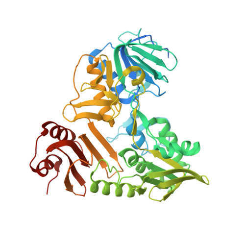
    5KVH, 5KVI

  • PubMed Abstract: 

    Apoptosis-inducing factor (AIF) is critical for mitochondrial respiratory complex biogenesis and for mediating necroptotic parthanatos; these functions are seemingly regulated by enigmatic allosteric switching driven by NADH charge-transfer complex (CTC) formation. Here, we define molecular pathways linking AIF's active site to allosteric switching regions by characterizing dimer-permissive mutants using small-angle X-ray scattering (SAXS) and crystallography and by probing AIF-CTC communication networks using molecular dynamics simulations. Collective results identify two pathways propagating allostery from the CTC active site: (1) active-site H454 links to S480 of AIF's central β-strand to modulate a hydrophobic border at the dimerization interface, and (2) an interaction network links AIF's FAD cofactor, central β-strand, and Cβ-clasp whereby R529 reorientation initiates C-loop release during CTC formation. This knowledge of AIF allostery and its flavoswitch mechanism provides a foundation for biologically understanding and biomedically controlling its participation in mitochondrial homeostasis and cell death.


  • Organizational Affiliation

    Biochemistry and Molecular Biophysics, Washington University School of Medicine, St. Louis, MO 63110, USA; Molecular and Cellular Oncology, The University of Texas M.D. Anderson Cancer Center, Houston, TX 77030, USA. Electronic address: cabrosey@mdanderson.org.


Macromolecules
Find similar proteins by:  (by identity cutoff)  |  3D Structure
Entity ID: 1
MoleculeChains Sequence LengthOrganismDetailsImage
Apoptosis-inducing factor 1, mitochondrial
A, B
543Homo sapiensMutation(s): 1 
Gene Names: AIFM1AIFPDCD8
EC: 1.1.1 (PDB Primary Data), 1.6.99 (UniProt)
UniProt & NIH Common Fund Data Resources
Find proteins for O95831 (Homo sapiens)
Explore O95831 
Go to UniProtKB:  O95831
PHAROS:  O95831
GTEx:  ENSG00000156709 
Entity Groups  
Sequence Clusters30% Identity50% Identity70% Identity90% Identity95% Identity100% Identity
UniProt GroupO95831
Sequence Annotations
Expand
  • Reference Sequence
Experimental Data & Validation

Experimental Data

  • Method: X-RAY DIFFRACTION
  • Resolution: 2.27 Å
  • R-Value Free:  0.239 (Depositor), 0.240 (DCC) 
  • R-Value Work:  0.202 (Depositor), 0.210 (DCC) 
  • R-Value Observed: 0.204 (Depositor) 
Space Group: P 21 21 21
Unit Cell:
Length ( Å )Angle ( ˚ )
a = 89.845α = 90
b = 112.166β = 90
c = 122.211γ = 90
Software Package:
Software NamePurpose
PHENIXrefinement
XDSdata reduction
Aimlessdata scaling
PHASERphasing

Structure Validation

View Full Validation Report


Ligand Structure Quality Assessment 

Created with Raphaël 2.3.0Worse 01 BetterLigand structure goodness of fit to experimental dataBest fitted FADClick on this verticalbar to view details

Entry History & Funding Information

Deposition Data


Funding OrganizationLocationGrant Number
National Institutes of Health/National Cancer Institute (NIH/NCI)United StatesP01-CA92584
National Institutes of Health/National Institute of General Medical Sciences (NIH/NIGMS)United StatesF32-GM109591

Revision History  (Full details and data files)

  • Version 1.0: 2016-11-16
    Type: Initial release
  • Version 1.1: 2016-12-14
    Changes: Database references
  • Version 1.2: 2017-09-06
    Changes: Author supporting evidence
  • Version 1.3: 2019-12-04
    Changes: Author supporting evidence
  • Version 1.4: 2023-10-04
    Changes: Data collection, Database references, Refinement description